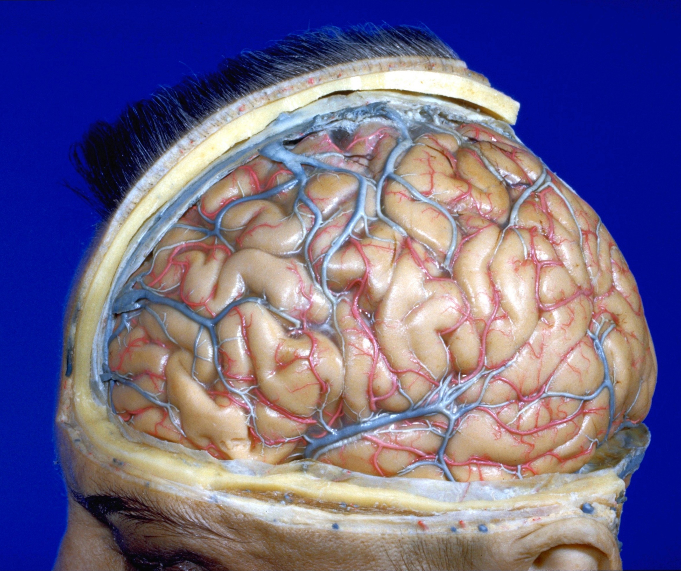
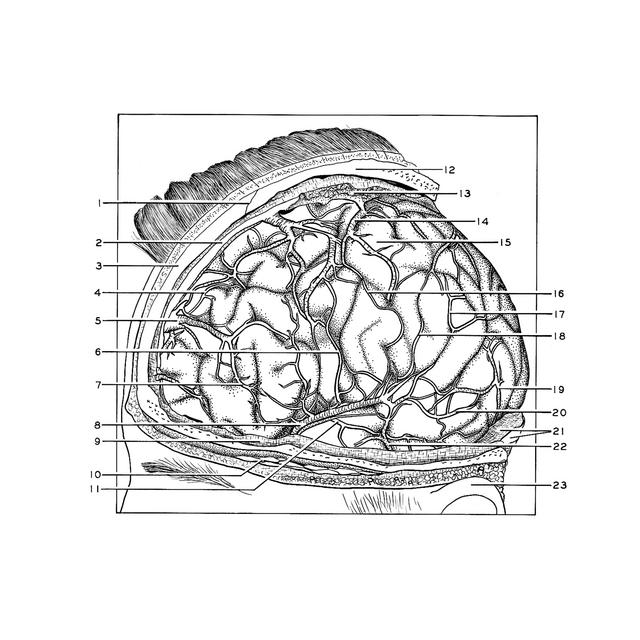
Exploration of the meninges and brain in situ
Dura removed, arachnoid membrane intact, cerebral veins and cortical arteries
Stanford holds the copyright to the David L. Bassett anatomical images and has assigned
Creative Commons license Attribution-Share
Alike 4.0 International to all of the images.
For additional information regarding use and permissions,
please contact Dr. Drew Bourn at dbourn@stanford.edu.
Image #1-4
Exploration of the meninges and brain in situ
Dura removed, arachnoid membrane intact, cerebral veins and cortical arteries
The dura mater has been cut away so that the left cerebral hemisphere is visible lying inside the arachnoid membrane. The tentorium appears in the lower right part of the view and the transverse sinus is opened along its posterior border. The superior sagittal sinus has been opened by removing a strip of dura from its superficial wall. The superior cerebral veins ascend on the surface of the frontal and parietal lobes to empty into this sinus at various points. A number of anastomoses are present between these veins, none being particularly large in this specimen. In general the superior cerebral veins are divided into anterior and posterior groups. In this case there appear to be several anterior vessels, a group of large veins intermediate in position (overlying the region of the central sulcus) and several posterior veins (not visible in this view).
- Coronal suture
- Superior sagittal sinus
- Frontal bone
- Branch anterior cerebral artery
- Superior cerebral vein (anterior)
- Artery of precentral sulcus
- Anastomotic connection between anterior superior cerebral vein and middle cerebral vein
- Middle cerebral vein
- Dura mater
- Temporalis muscle
- Anterior temporal branch of middle cerebral artery
- Parietal bone
- Venous lacuna
- Superior cerebral vein (overlying central sulcus)
- Postcentral gyrus
- Artery of central sulcus
- Parietal branch of middle cerebral artery
- Artery of postcentral sulcus
- Inferior cerebral vein (lateral occipital vein)
- Posterior temporal branch of middle cerebral artery
- Tentorium cerebelli and transverse sinus
- Small anastomotic vein of Labbé
- Auricle