Bassett Collection of Stereoscopic Images of Human Anatomy
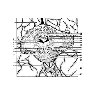
Serial transverse sections of the brain stem
Junction of medulla and pons.
Image #30-2
KEYWORDS: Brain, Cerebellum, Medulla, Midbrain, Pons, Ventricules.
Creative Commons
Stanford holds the copyright to the David L. Bassett anatomical images and has assigned Creative Commons license Attribution-Share Alike 4.0 International to all of the images.

For additional information regarding use and permissions, please contact Dr. Drew Bourn at dbourn@stanford.edu.

|
| ||||||||||||||||||||||||||||||||||||||||||||||||||||||||||

|
|