Bassett Collection of Stereoscopic Images of Human Anatomy
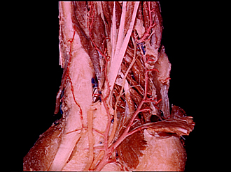
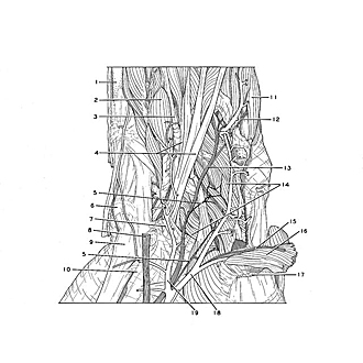
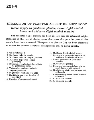
Dissection of plantar aspect of left foot
Nerve supply to quadratus plantae, flexor digiti minimi brevis and abductor digiti minimi muscles
Image #201-4
KEYWORDS: Foot and toes, Muscles and tendons, Peripheral nervous system.
Creative Commons
Stanford holds the copyright to the David L. Bassett anatomical images and has assigned Creative Commons license Attribution-Share Alike 4.0 International to all of the images.

For additional information regarding use and permissions, please contact Dr. Drew Bourn at dbourn@stanford.edu.
|
| ||||||||||||||||||||||||||||||||||||||||||
|
|