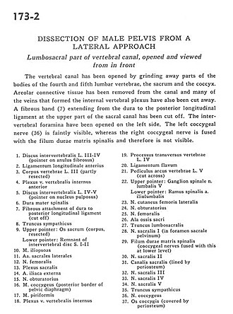
| 1
.
| Intervertebral disc L. III-IV (pointer on anulus fibrosus) |
| 2
.
| Anterior longitudinal ligament |
| 3
.
| Body of vertebra L. III (partly resected) |
| 4
.
| Anterior internal vertebral venous plexus |
| 5
.
| Intervertebral disc L. IV-V (pointer on nucleus pulposus) |
| 6
.
| Spinal dura mater |
| 7
.
| Fibrous attachment of dura to posterior longitudinal ligament (cut off) |
| 8
.
| Sympathetic trunk |
| 9
.
| Upper pointer: Sacrum (corpus, resected) Lower pointer: Remnant of intervertebral disc S. I-lI |
| 10
.
| Iliopsoas muscle |
| 11
.
| Lateral sacral arteries |
| 12
.
| Femoral nerve |
| 13
.
| Sacral plexus |
| 14
.
| External iliac artery |
| 15
.
| Obturator nerve |
| 16
.
| Coccygeus muscle (posterior border of pelvic diaphragm) |
| 17
.
| Piriform muscle |
| 18
.
| Internal vertebral venous plexus |
| 19
.
| Transverse process vertebrae L. IV |
| 20
.
| Ligamentum flavum |
| 21
.
| Pedicle of arch of vertebra L. V (cut across) |
| 22
.
| Upper pointer: Spinal ganglion lumbar nerve V Lower pointer: Spinal branch iliolumbar artery |
| 23
.
| Lateral femoral cutaneous nerve |
| 24
.
| Obturator nerve |
| 25
.
| Femoral nerve |
| 26
.
| Ala of sacrum |
| 27
.
| Lumbosacral trunk |
| 28
.
| Sacral nerve I (in anterior (pelvic) sacral foramen) |
| 29
.
| Dural filum spinal cord (coccygeal nerves fused with this at lower level) |
| 30
.
| Sacral nerve II |
| 31
.
| Sacral canal (lined by periosteum) |
| 32
.
| Sacral nerve III |
| 33
.
| Sacral nerve IV |
| 34
.
| Sacral nerve V |
| 35
.
| Sympathetic trunk |
| 36
.
| Coccygeal nerve |
| 37
.
| Coccyx (covered by periosteum) |