Bassett Collection of Stereoscopic Images of Human Anatomy
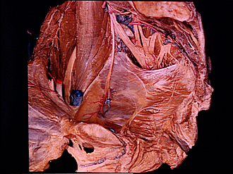
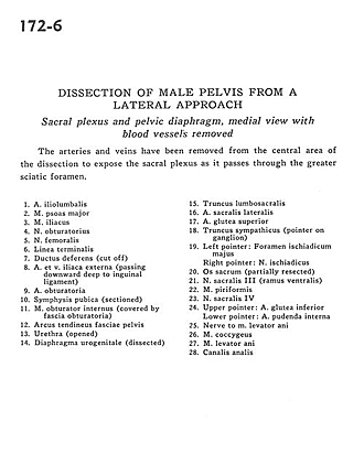
Dissection of male pelvis from a lateral approach
Sacral plexus and pelvic diaphragm, medial view with blood vessels removed
Image #172-6
KEYWORDS: Muscles and tendons, Vasculature.
Creative Commons
Stanford holds the copyright to the David L. Bassett anatomical images and has assigned Creative Commons license Attribution-Share Alike 4.0 International to all of the images.

For additional information regarding use and permissions, please contact Dr. Drew Bourn at dbourn@stanford.edu.
|

| ||||||||||||||||||||||||||||||||||||||||||||||||||||||||||||
|
|