| 1
.
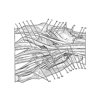
| Sheath ligament (cut to release enclosed flexor tendons) |
| 2
.
| Deep transverse metacarpal ligament |
| 3
.
| Flexor digitorum superficialis (tendons cut off) |
| 4
.
| Dorsal interosseous muscle I |
| 5
.
| Adductor pollicis muscle |
| 6
.
| Ligament of digital sheath |
| 7
.
| Abductor pollicis brevis muscle |
| 8
.
| Flexor pollicis brevis muscle (deep head) |
| 9
.
| Flexor pollicis longus muscle (tendon of insertion) |
| 10
.
| Muscular slip originating with flexor pollicis brevis muscle and inserting with opponens pollicis muscle |
| 11
.
| Opponens pollicis muscle |
| 12
.
| Abductor pollicis longus muscle (tendons of insertion) |
| 13
.
| Flexor carpi radialis muscle (tendon of insertion) |
| 14
.
| Superficial anterior branch radial artery |
| 15
.
| Flexor carpi ulnaris muscle (tendon of insertion) |
| 16
.
| Ulnar artery |
| 17
.
| Deep surface of carpal tunnel |
| 18
.
| Flexor digitorum profundus muscle |
| 19
.
| Deep branch of ulnar nerve (terminal branch) |
| 20
.
| Opponens digiti minimi muscle |
| 21
.
| Flexor digiti minimi muscle |
| 22
.
| Lumbrical muscles |
| 23
.
| Tendons of flexor digitorum profundus muscle |
| 24
.
| Aberrant slips of lumbrical muscles III-IV |