Arm
Right biceps muscle. anterior view
Stanford holds the copyright to the David L. Bassett anatomical images and has assigned
Creative Commons license Attribution-Share
Alike 4.0 International to all of the images.
For additional information regarding use and permissions,
please contact the Medical History Center.
Image #95-3
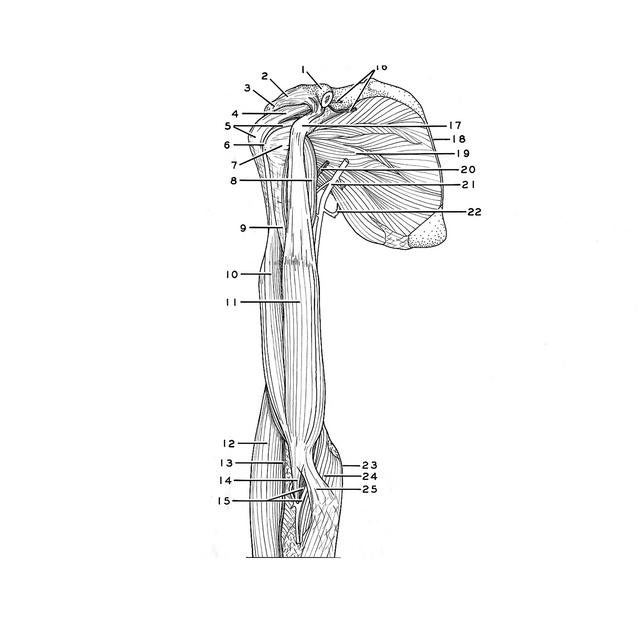
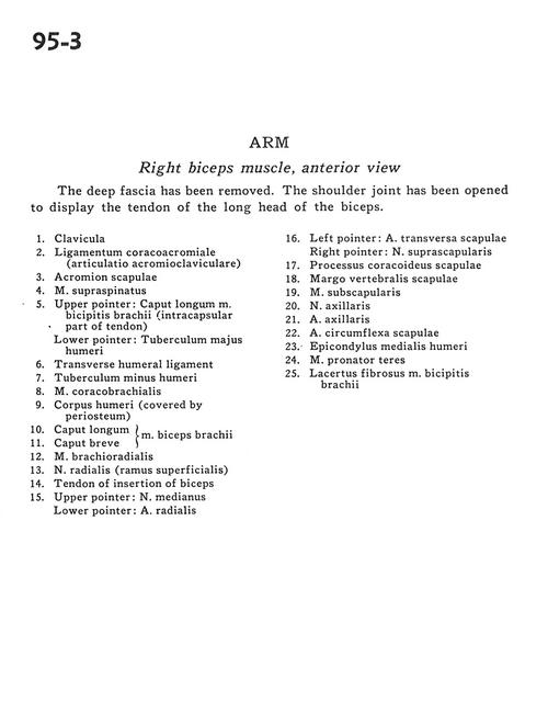
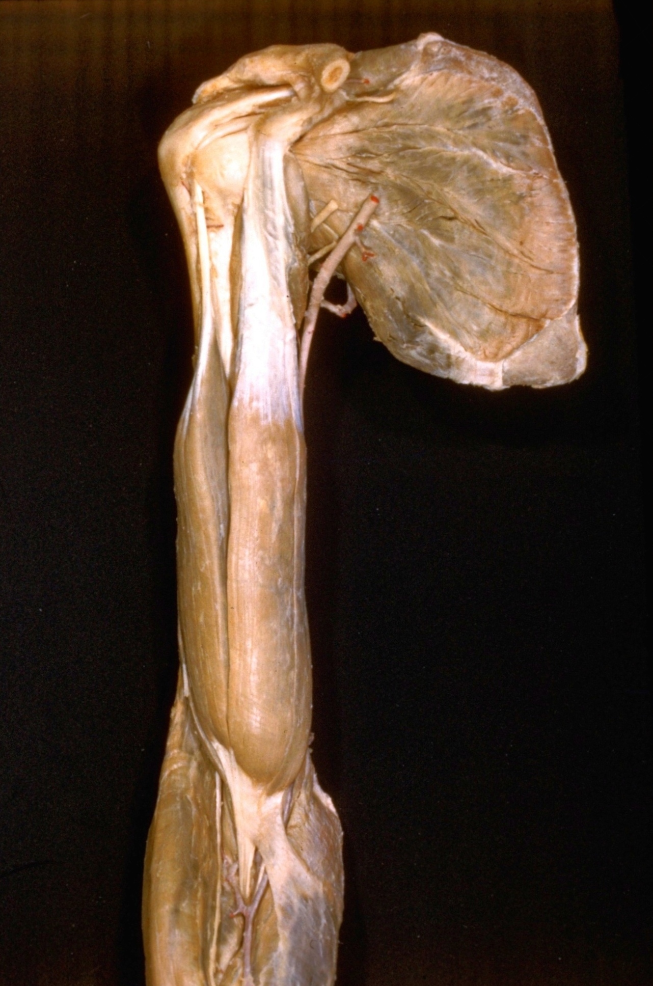
Arm
Right biceps muscle. anterior view
The deep fascia has been removed. The shoulder joint has been opened to display the tendon of the long head of the biceps.
- Clavicle
- Coracoacromial ligament (acromioclavicular joint)
- Acromion of scapula
- Supraspinatus muscle
- Upper pointer: Long head of biceps brachii muscle (intracapsular part of tendon) Lower pointer: Greater tubercle of humerus
- Transverse humeral ligament
- Lesser tubercle of humerus
- Coracobrachialis muscle
- Body of humerus (covered by periosteum)
- Long head biceps brachii muscle
- Short head
- Brachioradialis muscle
- Radial nerve (superficial branch)
- Tendon of insertion of biceps
- Upper pointer: Median nerve Lower pointer: Radial artery
- Left pointer: Transverse scapular artery Right pointer: Suprascapular nerve
- Coracoid process of scapula
- Vertebral margin of scapula
- Subscapularis muscle
- Axillary nerve
- Axillary artery
- Circumflex artery of scapula
- Medial epicondyle of humerus
- Pronator teres muscle
- Bicipital aponeurosis