
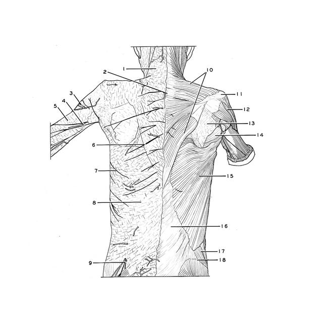
Shoulder
Right lattisimus dorsi and trapezius muscles, posterior view
Stanford holds the copyright to the David L. Bassett anatomical images and has assigned
Creative Commons license Attribution-Share
Alike 4.0 International to all of the images.
For additional information regarding use and permissions,
please contact the Medical History Center.
Image #92-6
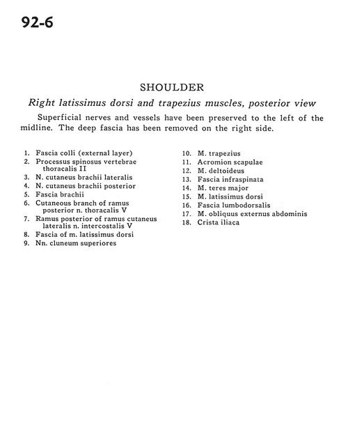
Shoulder
Right lattisimus dorsi and trapezius muscles, posterior view
Superficial nerves and vessels have been preserved to the left of the midline. The deep fascia has been removed on the right side.
- Superficial fascia (external layer)
- Spinous process thoracic vertebra II
- Lateral brachial cutaneous
- Posterior brachial cutaneous nerve
- Brachial fascia
- Cutaneous branch of branch posterior thoracic nerve V
- Posterior branch of lateral cutaneous branch of intercostal nerve V
- Fascia of latissimus dorsi muscle
- Superior cluneal nerves
- Trapezius muscle
- Acromion of scapula
- Deltoid muscle
- Infraspinatus fascia
- Teres major muscle
- Latissimus dorsi muscle
- Lumbodorsal fascia
- External abdominal oblique muscle
- Hiac crest