Pectoral region and axilla
Left sternoclavicular joint capsule and ligaments
Stanford holds the copyright to the David L. Bassett anatomical images and has assigned
Creative Commons license Attribution-Share
Alike 4.0 International to all of the images.
For additional information regarding use and permissions,
please contact the Medical History Center.
Image #90-7
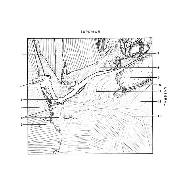
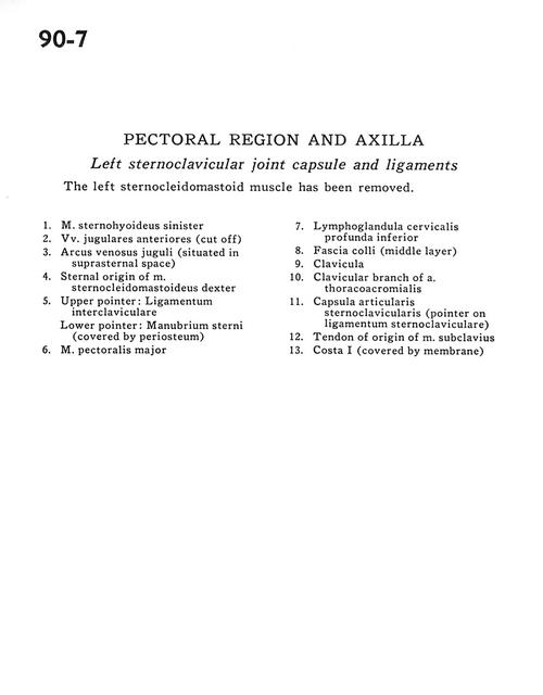
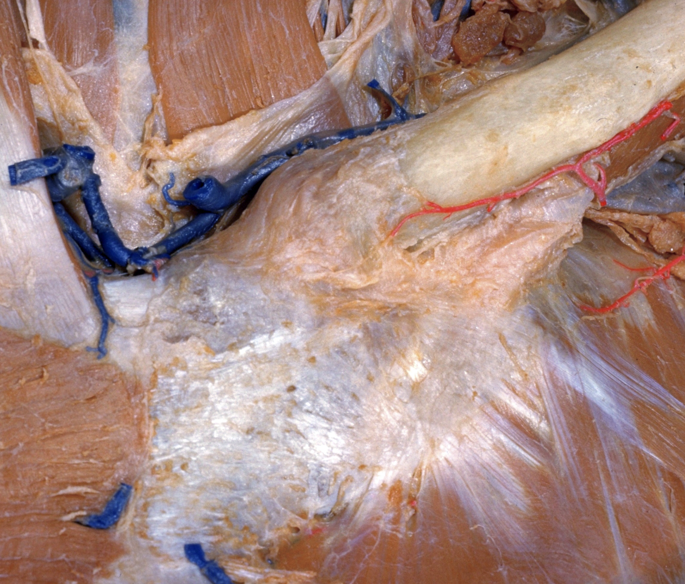
Pectoral region and axilla
Left sternoclavicular joint capsule and ligaments
The left sternocleidomastoid muscle has been removed.
- Left sternohyoid muscle
- Anterior jugular veins (cut off)
- Jugular venous arch (situated in suprasternal space)
- Sternal origin of sternocleidomastoid muscle
- Upper pointer: Interclavicular ligament Lower pointer: Manubrium of sternum (covered by periosteum)
- Pectoralis major muscle
- Inferior deep cervical lymph node
- Superficial fascia (middle layer)
- Clavicle
- Clavicular branch of thoracoacromial artery
- Sternoclavicular joint capsule (pointer on sternoclavicular ligament)
- Tendon of origin of subclavius muscle
- Rib I (covered by membrane)