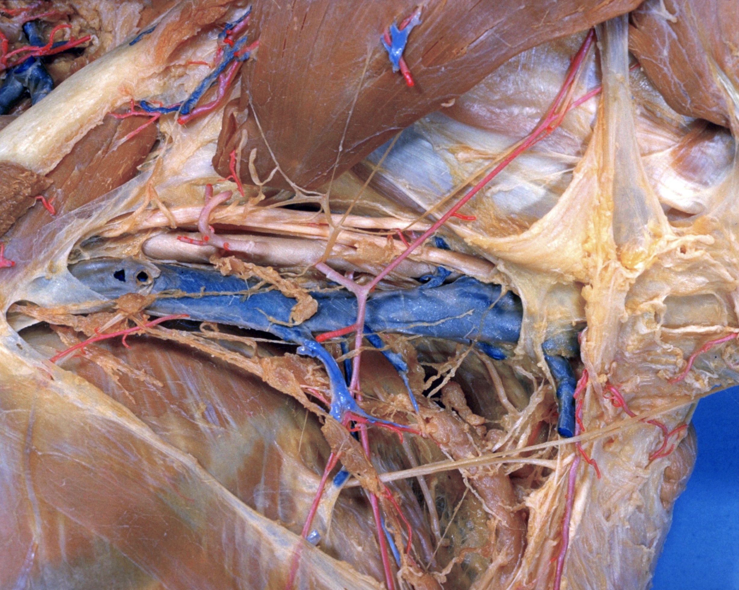
Pectoral region and axilla
Left axilla (continued); relations of vessels, nerves and lymphatic structures, close-up view
Stanford holds the copyright to the David L. Bassett anatomical images and has assigned
Creative Commons license Attribution-Share
Alike 4.0 International to all of the images.
For additional information regarding use and permissions,
please contact the Medical History Center.
Image #90-3
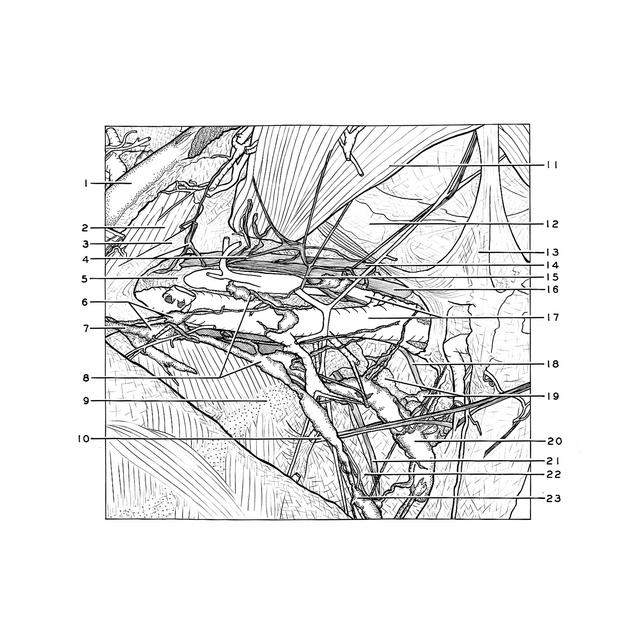
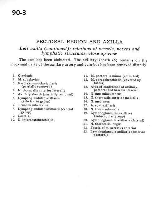
Pectoral region and axilla
Left axilla (continued); relations of vessels, nerves and lymphatic structures, close-up view
The arm has been abducted. The axillary sheath (5) remains on the proximal parts of the axillary artery and vein but has been removed distally.
- Clavicle
- Subclavius muscle
- Coracoclavicular fascia (partially removed)
- Anterior lateral thoracic nerve
- Axillary sheath (partially removed)
- Axillary lymph nodes (subclavian group)
- Subclavian trunk
- Axillary lymph nodes (central group)
- Rib II
- Intercostobrachial nerve
- Pectoralis minor muscle (reflected)
- Coracobrachialis muscle (covered by fascia)
- Area of confluence of axillary, pectoral and brachial fascia
- Musculocutaneous nerve
- Anterior medial thoracic nerve
- Median nerve
- Axillary artery and vein
- Thoracodorsal nerve
- Axillary lymph nodes (subscapular group)
- Axillary lymph node (lateral)
- Long thoracic nerve
- Fascia of serratus anterior muscle
- Axillary lymph node (anterior pectoral)