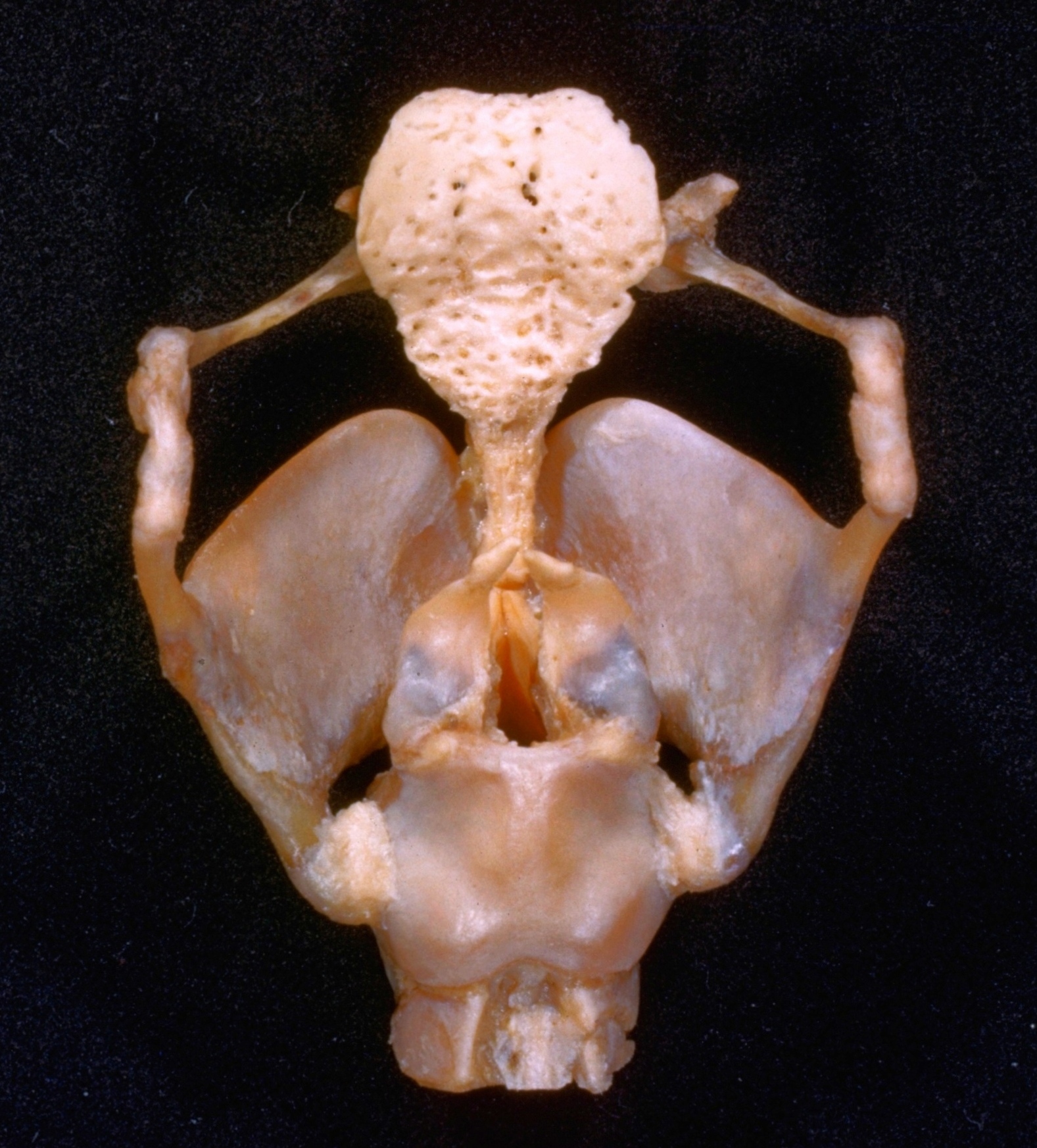
Hyoid bone and framework of larynx
Posterior view
Stanford holds the copyright to the David L. Bassett anatomical images and has assigned
Creative Commons license Attribution-Share
Alike 4.0 International to all of the images.
For additional information regarding use and permissions,
please contact the Medical History Center.
Image #84-2
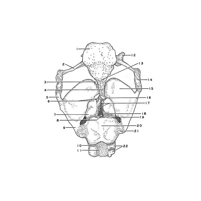
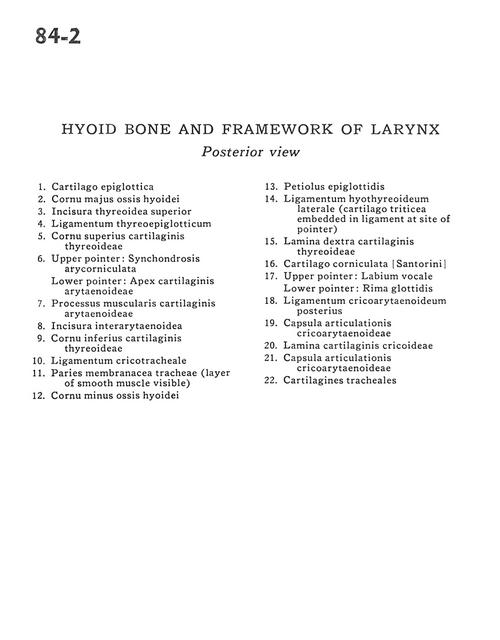
Hyoid bone and framework of larynx
Posterior view
- Epiglottic cartilage
- Greater horn hyoid bone
- Superior thyroid incisure
- Thyroepiglottic ligament
- Superior horn thyroid cartilage
- Upper pointer: Arycorniculate synchondrosis Lower pointer: Apex of arytenoid cartilage
- Muscular process of arytenoid cartilage
- Interarytenoid notch
- Inferior horn of thyroid cartilage
- Cricotracheal ligament
- Membranous wall of trachea (layer of smooth muscle visible)
- Lesser horn hyoid bone
- Stem of epiglottis
- Lateral thyrohyoid ligament (Triticeal cartilage embedded in ligament at site of pointer)
- Lamina right thyroid cartilage
- Corniculate cartilage
- Upper pointer: Vocal lip Lower pointer: Rima glottidis
- Posterior cricoarytenoid ligament
- Cricoarytenoid joint capsule
- Lamina of cricoid cartilage
- Cricoarytenoid joint capsule
- Tracheal cartilage