Dissection of head and neck from a posterior approach
Larynx; posterior surface view
Stanford holds the copyright to the David L. Bassett anatomical images and has assigned
Creative Commons license Attribution-Share
Alike 4.0 International to all of the images.
For additional information regarding use and permissions,
please contact the Medical History Center.
Image #83-2
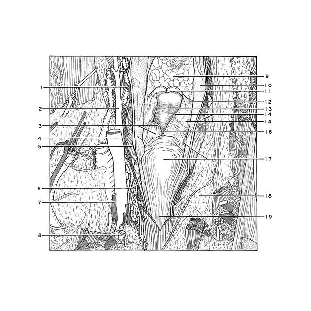
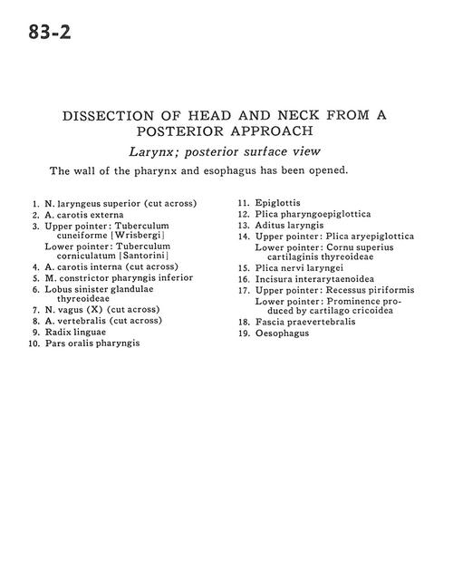
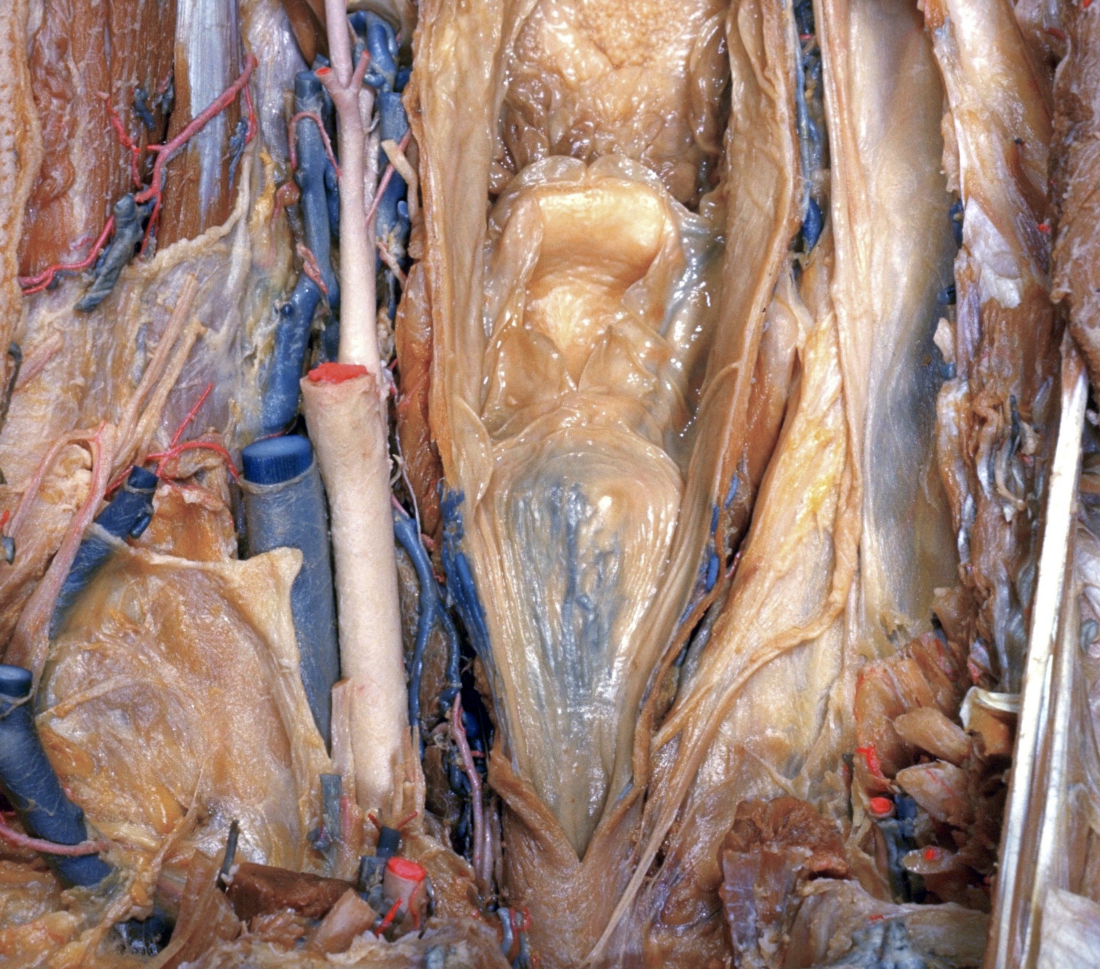
Dissection of head and neck from a posterior approach
Larynx; posterior surface view
The wall of the pharynx and esophagus has been opened.
- Superior laryngeal nerve (cut across)
- External carotid artery
- Upper pointer: Cuneiform tubercle Lower pointer: Corniculate tubercle
- Internal carotid artery (cut across)
- Inferior pharyngeal constrictor muscle
- Left lobe of thyroid gland
- Vagus nerve (X) (cut across)
- Vertebral artery (cut across)
- Root of tongue
- Oral part pharynx
- Epiglottis
- Pharyngoepiglottic fold
- Laryngeal ventricle
- Upper pointer: Aryepiglottic fold Lower pointer: Superior horn thyroid cartilage
- Fold of laryngeal nerve
- Interarytenoid incisure
- Upper pointer: Piriform recess Lower pointer: Prominence produced by cricoid cartilage
- Prevertebral fascia
- Esophagus