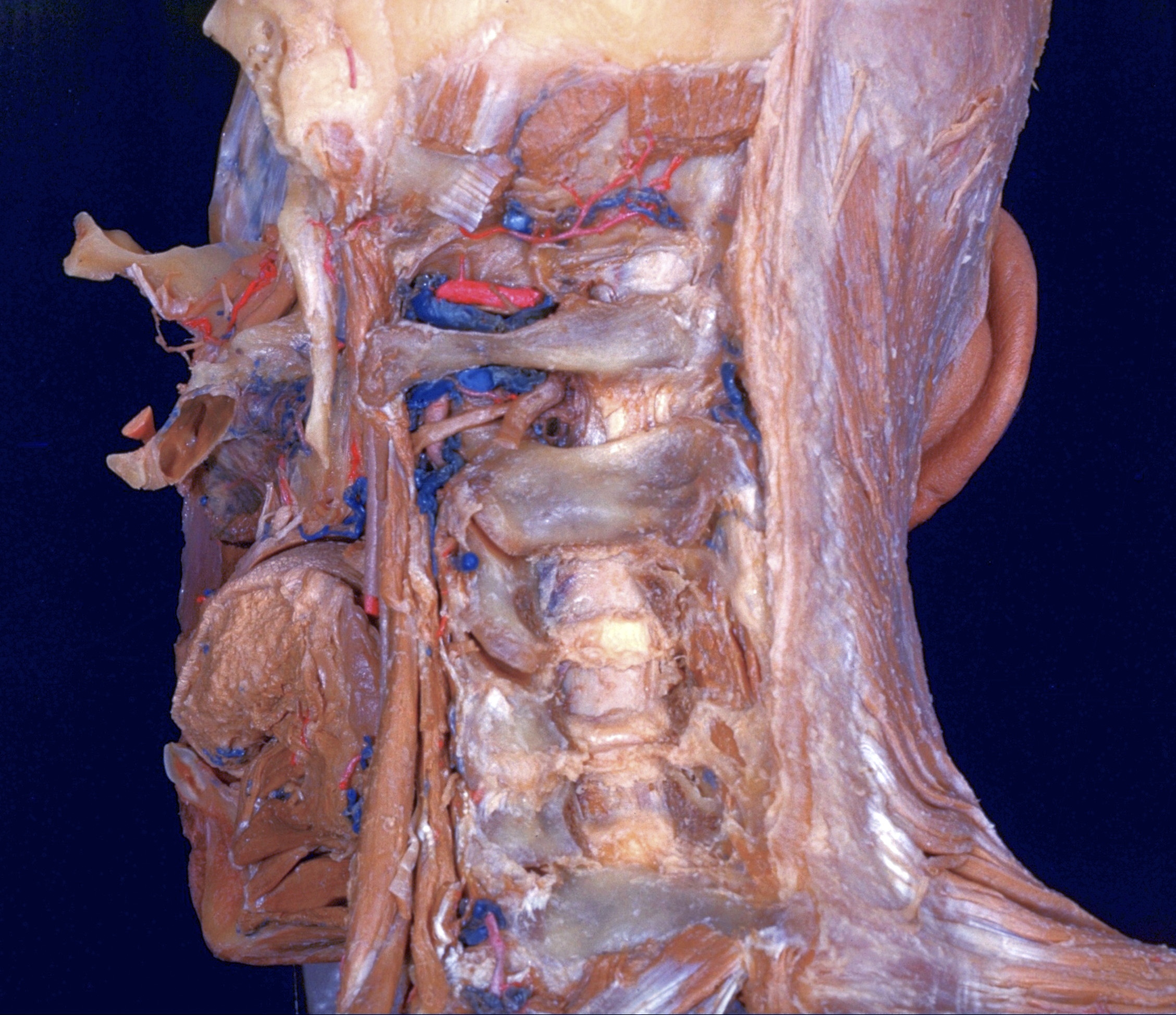
Dissection of head and neck from a posterior approach
Ligamenta flava
Stanford holds the copyright to the David L. Bassett anatomical images and has assigned
Creative Commons license Attribution-Share
Alike 4.0 International to all of the images.
For additional information regarding use and permissions,
please contact the Medical History Center.
Image #80-1
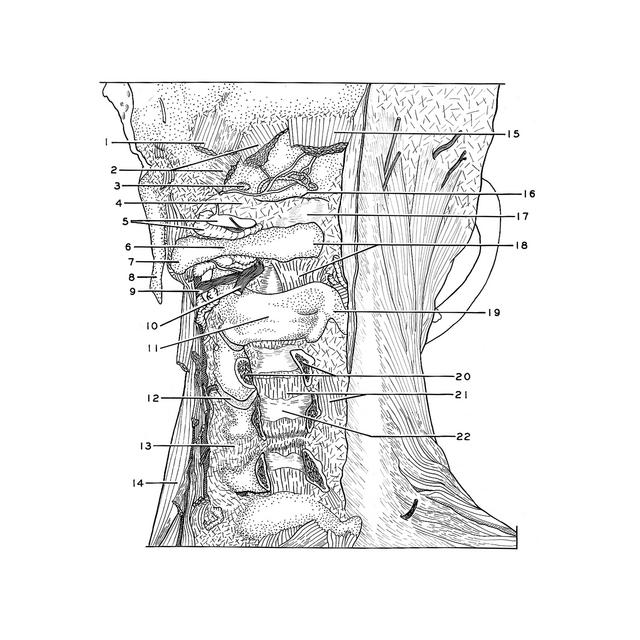
Dissection of head and neck from a posterior approach
Ligamenta flava
The arches of the third, fourth and fifth cervical vertebrae have been divided. The cavities of the superior and inferior (12) intervertebral joints of the third cervical vertebrae have been opened.
- Superior oblique capitis muscle (cut across)
- Major posterior rectus capitis muscle (cut across)
- Condyloid emissary
- Atlanto-occipital joint capsule
- Vertebral artery and vein
- Posterior arch of atlas
- Transverse process atlas
- Styloid process
- Anterior branch of cervical nerve II (note relation to vertebral artery)
- Greater occipital nerve
- Arch axis
- Articular cavity
- Joint capsule
- Middle scalene muscle
- Minor posterior rectus capitis muscle (cut across)
- Border of foramen magnum
- Dura mater
- Upper pointer: Posteror tubercle atlas Lower pointer: Ligamentum flavum
- Spinous process axis
- Cut margins of arch of cervical vertebra III (resected)
- Upper pointer: Ligamentum flavum Lower pointer: Interspinalis muscle
- Dura mater (epidural space absent at this level)