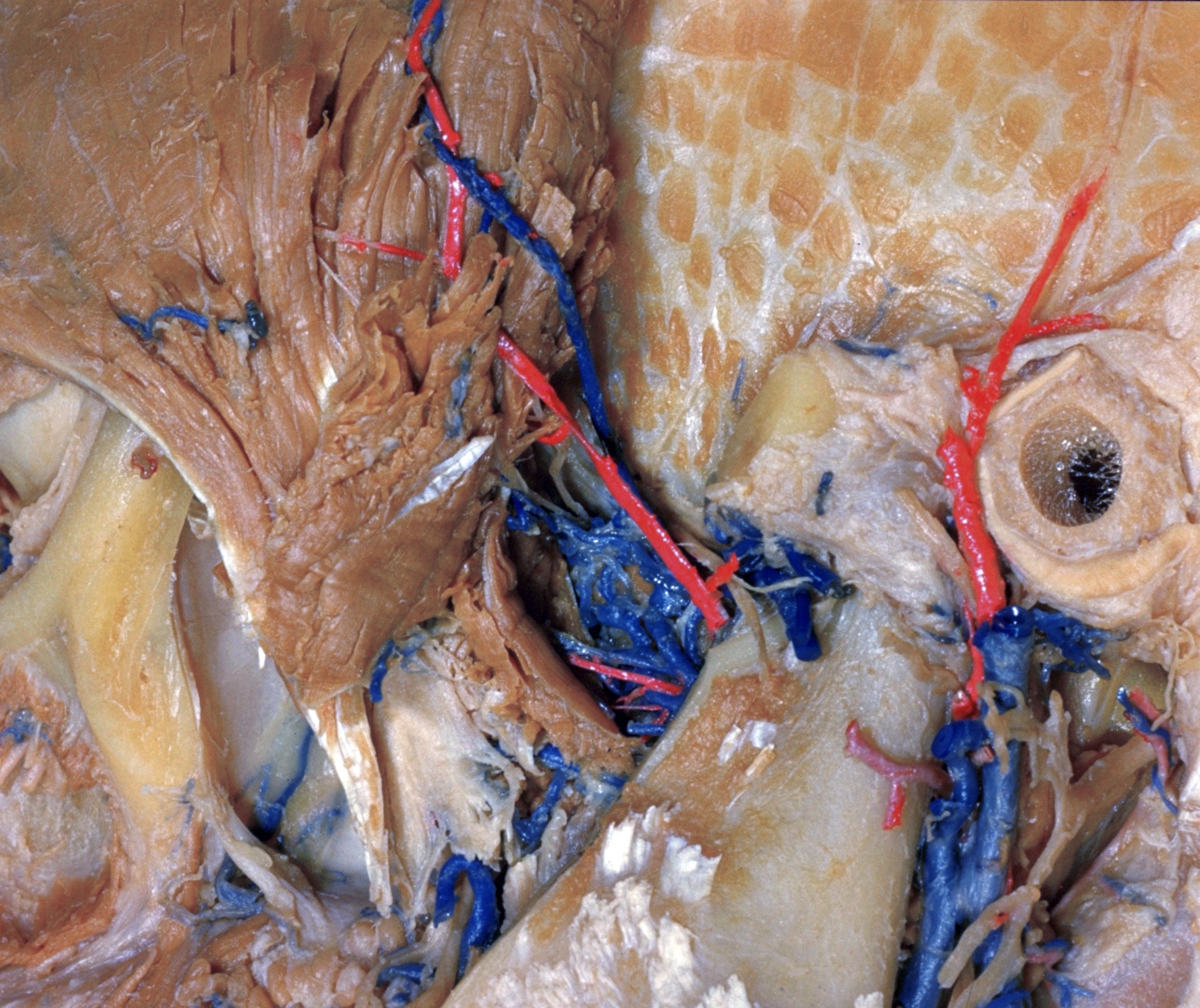
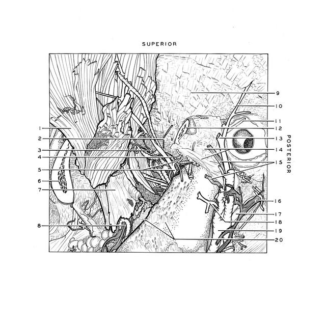
Dissection of left temporal region
Posterior deep temporal nerve and artery; capsule of temporomandibular joint, lateral view
Stanford holds the copyright to the David L. Bassett anatomical images and has assigned
Creative Commons license Attribution-Share
Alike 4.0 International to all of the images.
For additional information regarding use and permissions,
please contact the Medical History Center.
Image #65-1
Dissection of left temporal region
Posterior deep temporal nerve and artery; capsule of temporomandibular joint, lateral view
The temporal muscle (1) has been reflected anteriorly. Its tendon of insertion (7) remains in situ, although the coronoid process to which it attached has been cut away (20).
- Posterior border of temporalis muscle turned anteriorly
- Branch posterior deep temporal nerve (cut off)
- Posterior deep temporal nerve
- Masseteric nerve
- Upper pointer: Masseteric artery Lower pointer: Deep posterior temporal artery
- Zygomatic arch (divided)
- Tendon of temporalis muscle (note impression produced by coronoid process of mandible)
- Buccal nerve
- Temporal fossa
- Middle temporal artery
- Cartilaginous external acoustic meatus
- Zygomatic arch (cut across)
- Superficial temporal artery (cut off)
- Upper pointer: Joint capsule of mandible Lower pointer: Temporomandibular ligament
- Branches of auriculotemporal nerve
- Facial nerve (VII)
- Parotid gland (deep lobe)
- Transverse facial artery (cut off)
- Posterior facial vein
- Upper pointer: Ramus of mandible Lower pointer: Coronoid process (cut across)