Dissection of ear from lateral aspect
Right auricular cartilage, medial surface
Stanford holds the copyright to the David L. Bassett anatomical images and has assigned
Creative Commons license Attribution-Share
Alike 4.0 International to all of the images.
For additional information regarding use and permissions,
please contact the Medical History Center.
Image #59-6
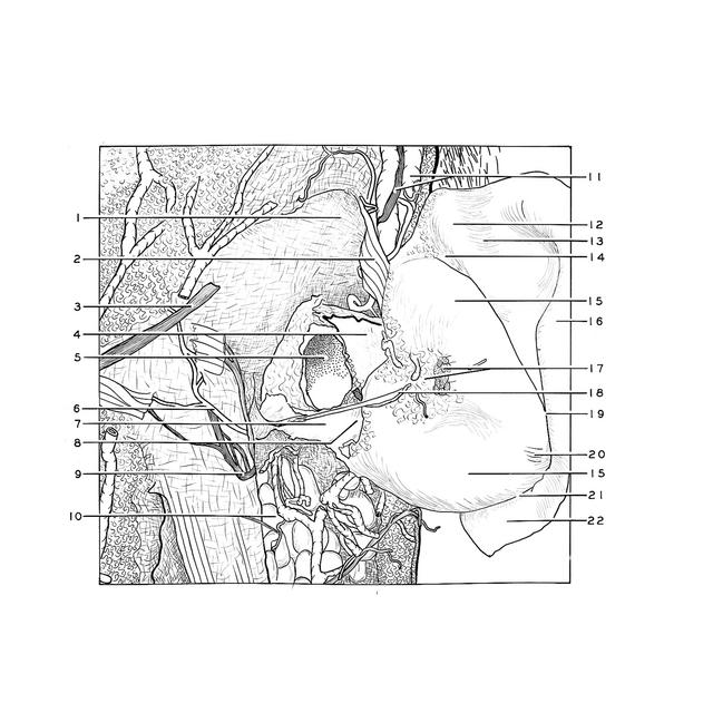

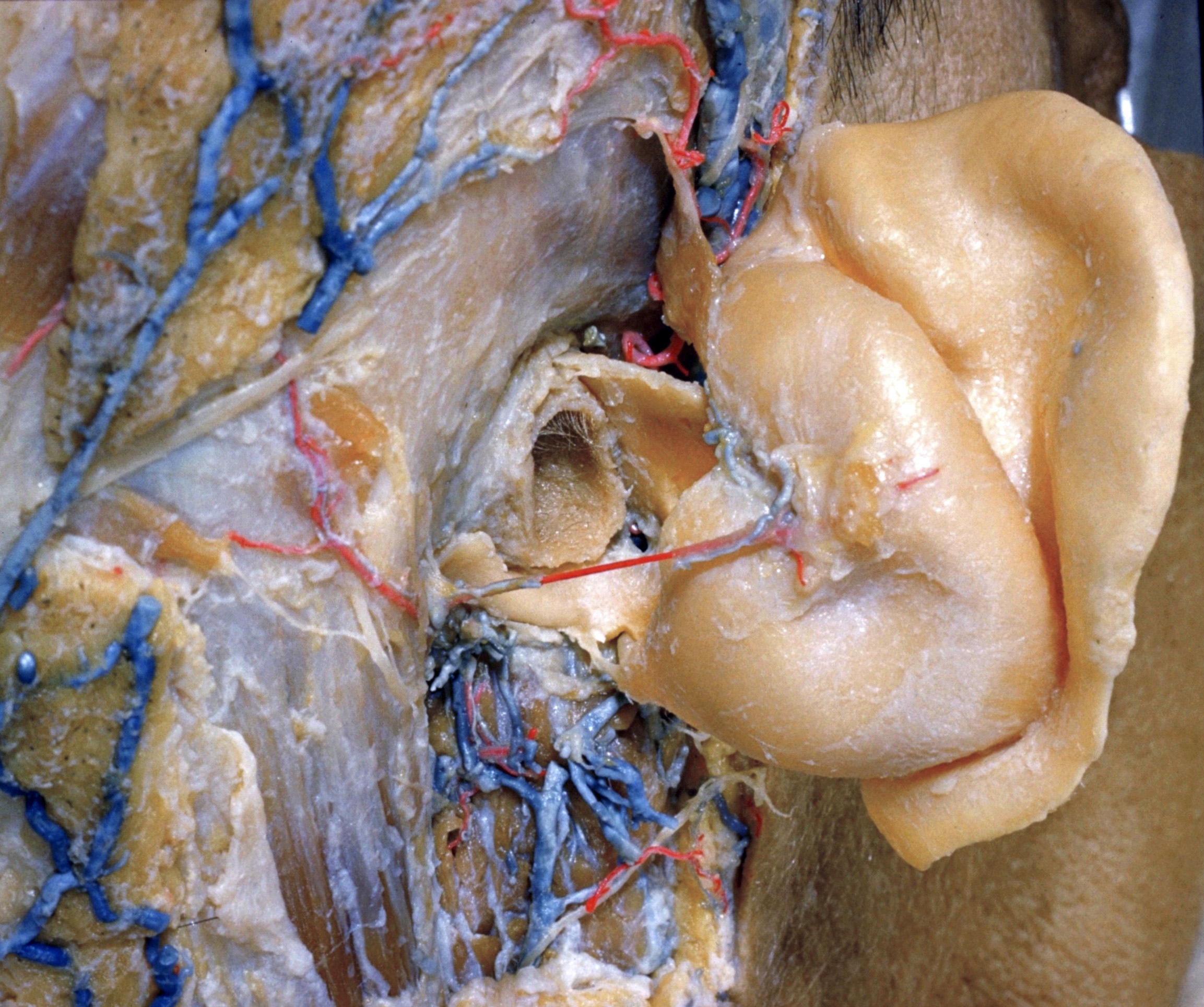
Dissection of ear from lateral aspect
Right auricular cartilage, medial surface
The cartilage has been retracted anterolaterally after removal of its perichondrium.
- Temporal fascia
- Superior auricular muscle
- Lesser occipital nerve
- Tragal plate
- External acoustic meatus
- Occipital branch posterior auricular artery
- Cartilaginous acoustic meatus
- Terminal incisure (pointer on cartilaginous isthmus)
- Posterior auricular nerve
- Parotid gland
- Superficial temporal vein and auriculotemporal nerve
- Eminence of triangular fossa
- Sulcus of upper crus of anthelix
- Transverse sulcus of anthelix
- Conchal eminence
- Scaphoid eminence
- Posterior auricular muscle and posterior auricular ligament (cut off)
- Posterior auricular branch auricular artery
- Fossa of anthelix
- Transverse auricular muscle
- Antitragicohelical fissure
- Tail of helix