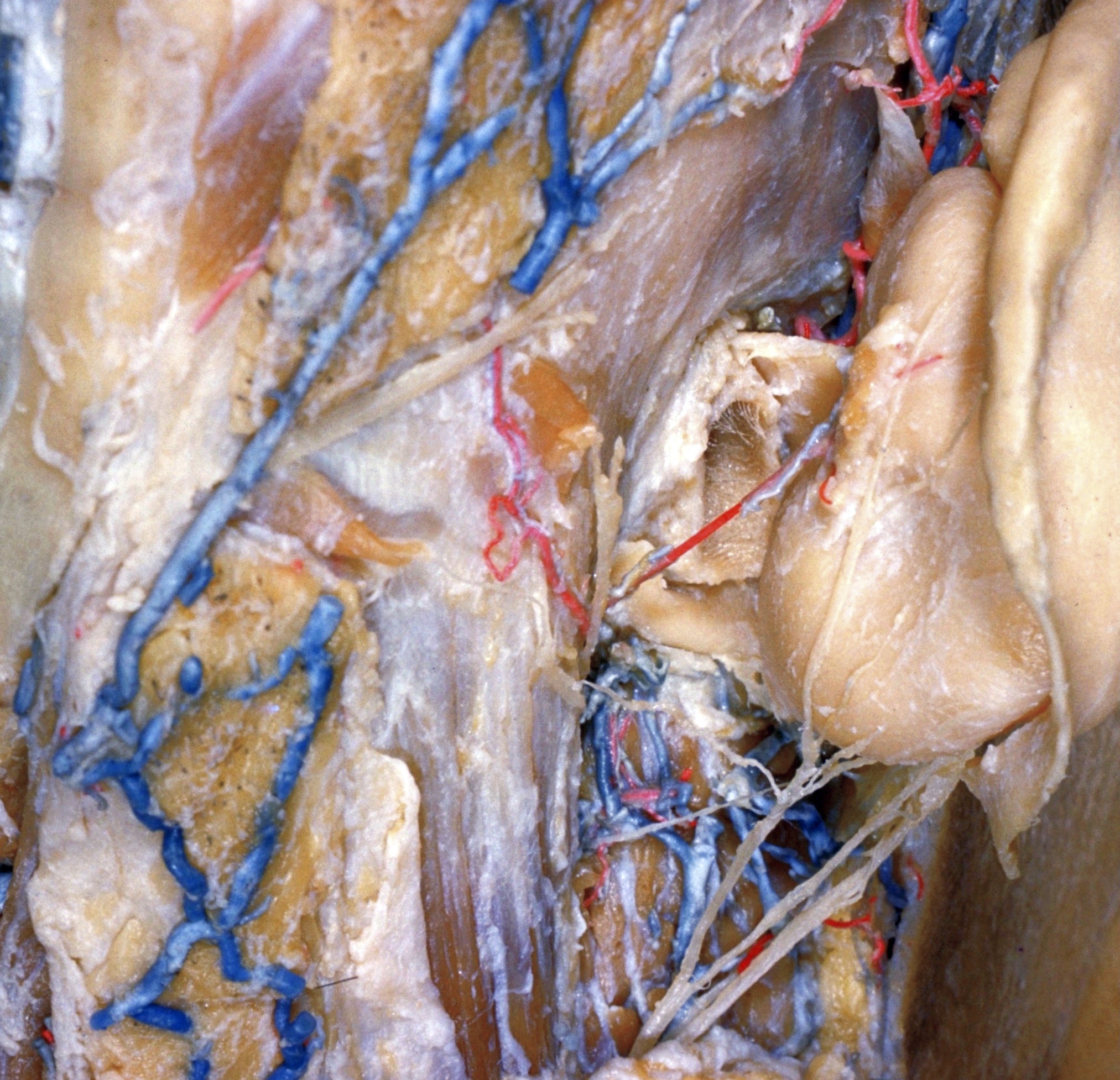
Dissection of ear from lateral aspect
Relation of right auricular cartilage to external auditory meatus; terminal branches of great auricular nerve
Stanford holds the copyright to the David L. Bassett anatomical images and has assigned
Creative Commons license Attribution-Share
Alike 4.0 International to all of the images.
For additional information regarding use and permissions,
please contact the Medical History Center.
Image #59-5
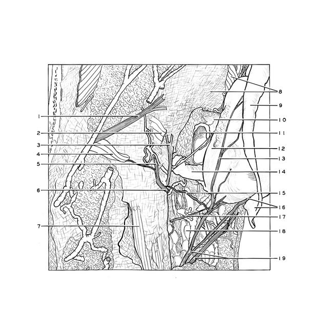
Dissection of ear from lateral aspect
Relation of right auricular cartilage to external auditory meatus; terminal branches of great auricular nerve
The posterior auricular muscle has been cut and the auricular cartilage retracted laterally.
- Lesser occipital nerve
- Posterior auricular muscle (cut across)
- Auricular branch vagus nerve
- Aberrant slip of posterior auricular muscle
- Occipital branch of posterior auricular nerve
- Posterior auricular nerve
- Sternocleidomastoid muscle (superficial fascia reflected)
- Upper pointer: Superior auricular muscle Lower pointer: Temporal fascia
- Helix
- Tragal plate
- External acoustic meatus (opened posteriorly)
- Conchal eminence
- Posterior branch greater auricular nerve
- Cartilaginous acoustic meatus
- Communication between posterior auricular nerve and greater auricular nerve
- Upper pointer: Antitragicohelical fissure Lower pointer: Tail of helix
- Branch facial nerve (to intrinsic muscles of auricle)
- Parotid gland
- Greater auricular nerve