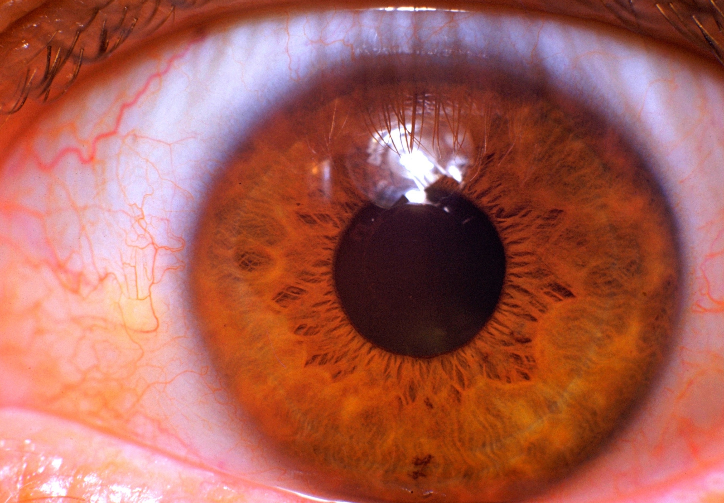
Dissection of eye
Right cornea, anterior view
Stanford holds the copyright to the David L. Bassett anatomical images and has assigned
Creative Commons license Attribution-Share
Alike 4.0 International to all of the images.
For additional information regarding use and permissions,
please contact the Medical History Center.
Image #58-4
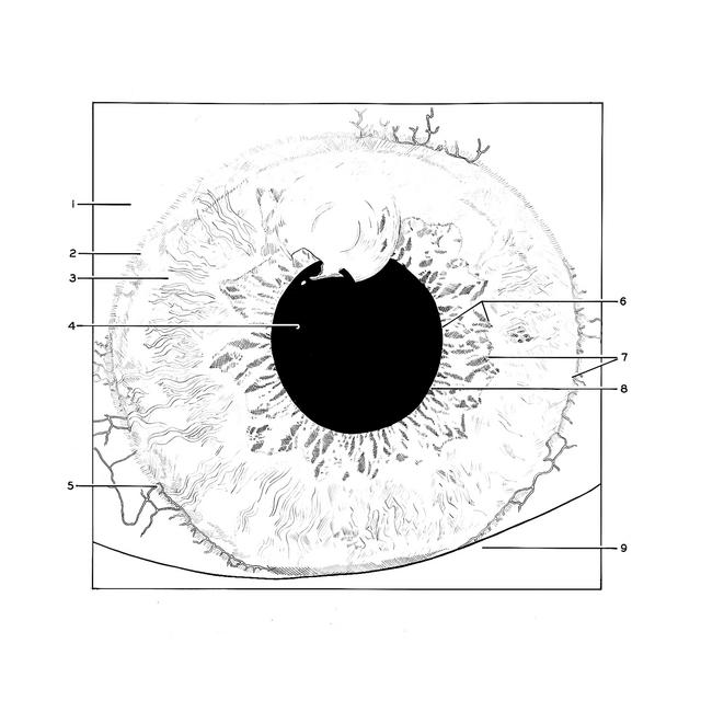
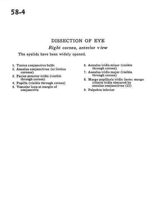
Dissection of eye
Right cornea, anterior view
The eyelids have been widely opened.
- Tunica bulbar conjunctiva
- Annulus conjunctiva (at limbus of cornea)
- Anterior surface of iris (visible through cornea)
- Pupil (visible through cornea)
- Vascular loop at margin of conjunctiva
- Annulus iris minor (visible through cornea)
- Annulus iris major (visible through cornea)
- Pupillary-iris margin (note: ciliary margin of iris obscured by annulus conjunctiva (2))
- Inferior palpebra