Floor of cranial cavity
Structures inferior to anterior and middle cranial fossae; orbit and sinuses opened
Stanford holds the copyright to the David L. Bassett anatomical images and has assigned
Creative Commons license Attribution-Share
Alike 4.0 International to all of the images.
For additional information regarding use and permissions,
please contact the Medical History Center.
Image #50-5
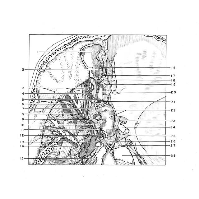
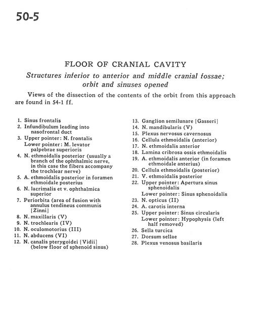
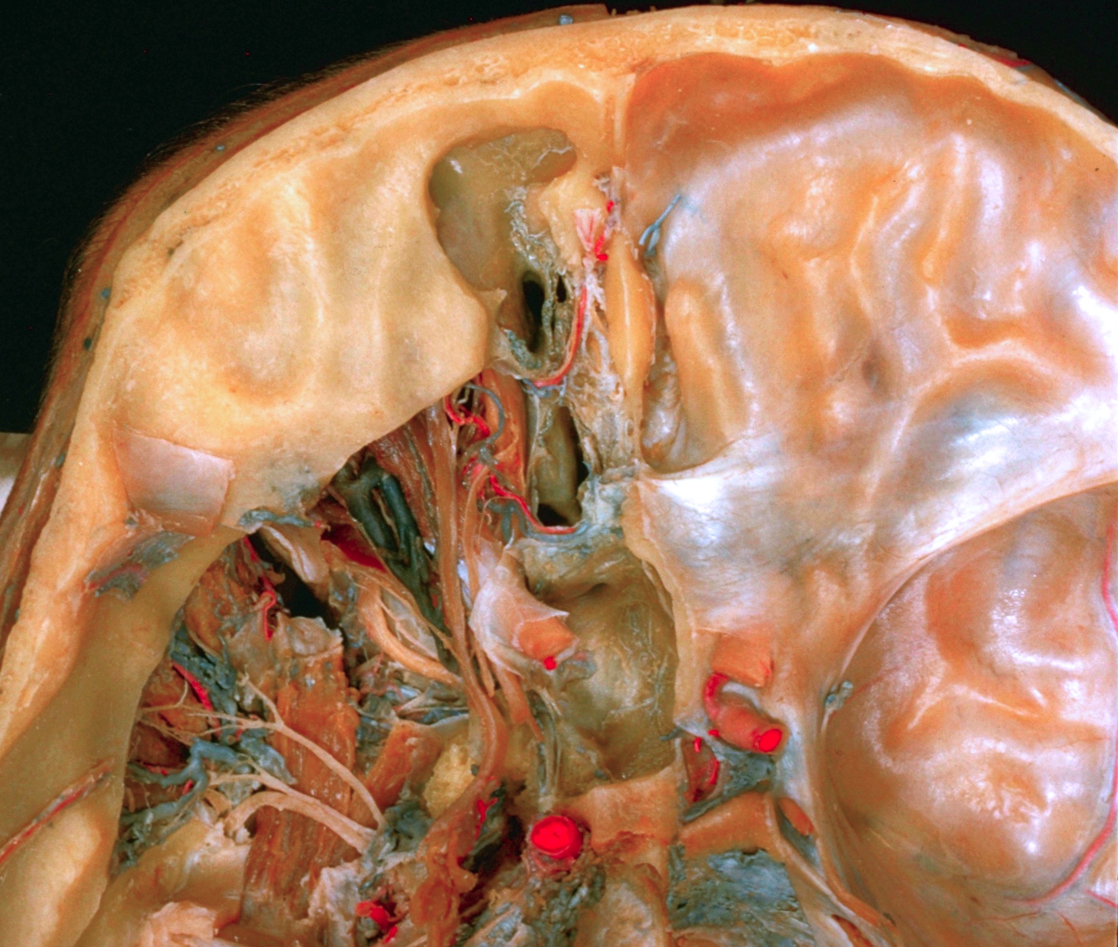
Floor of cranial cavity
Structures inferior to anterior and middle cranial fossae; orbit and sinuses opened
Views of the dissection of the contents of the orbit from this approach are found in 54-1 ff.
- Frontal sinus
- Infundibulum leading into nasofrontal duct
- Upper pointer: Frontal nerve Lower pointer: Levator palpebrae superioris
- Posterior ethmoidal nerve (usually a branch of the ophthalmic nerve in this case the fibers accompany the trochlear nerve)
- Posterior ethmoidal artery in posterior ethmoidal foramen
- Lacrimal nerve and superior ophthalmic vein
- Periorbita (area of fusion with common annular tendon)
- Maxillary nerve (V2)
- Trochlear nerve (IV)
- Oculomotor nerve (III)
- Abducens nerve (VI)
- Vidian nerve of pterygoid canal (below floor of sphenoid sinus)
- Semilunar ganglion (trigeminal)
- Mandibular nerve (V3)
- Cavernous nerve plexus
- Ethmoidal cell (anterior)
- Anterior ethmoidal nerve
- Cribriform plate ethmoid bone
- Anterior ethmoidal artery (in anterior ethmoidal foramen)
- Ethmoidal cell (posterior)
- Posterior ethmoidal vein
- Upper pointer: Aperture of sphenoid sinus Lower pointer: Sphenoid sinus
- Optic nerve (ll)
- Internal carotid artery
- Upper pointer: Circular sinus Lower pointer: Hypophysis (left half removed)
- Sella turcica
- Dorsum sellae
- Basilar venous plexus