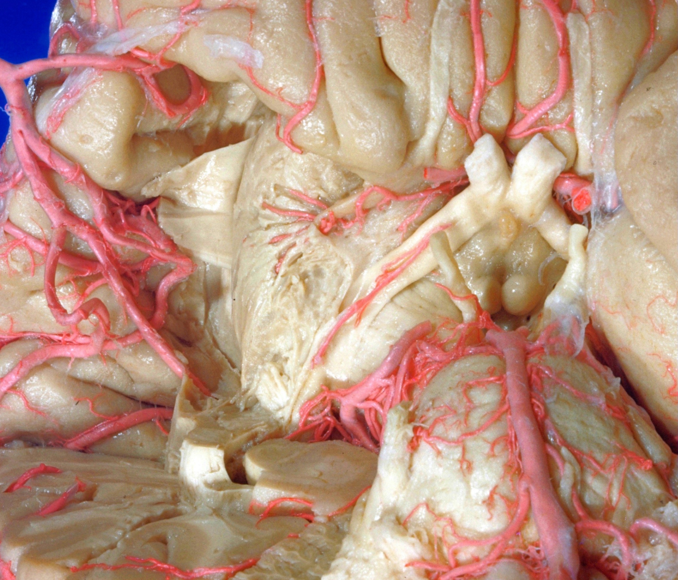
Exploration of the brain from its basal aspect
External capsule, lentiform nucleus and deeper course of striate arteries
Stanford holds the copyright to the David L. Bassett anatomical images and has assigned
Creative Commons license Attribution-Share
Alike 4.0 International to all of the images.
For additional information regarding use and permissions,
please contact the Medical History Center.
Image #5-5
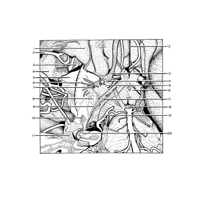
Exploration of the brain from its basal aspect
External capsule, lentiform nucleus and deeper course of striate arteries
The main trunk of the middle cerebral artery has been retracted far laterally, the medullary center of the insula removed and the claustrum scraped away. The external capsule has been partially removed, the remaining portion being turned laterally to expose the lentiform nucleus. The course of the lateral striate arteries into the lentiform nucleus is visible.
- Orbital gyrus
- Middle cerebral artery (divided and turned laterally)
- Superior longitudinal fasciculus
- Frontal part of operculum
- External capsule (reflected laterally)
- Lentiform nucleus (putamen)
- Lateral striate artery
- Amygdaloid nucleus (partially removed)
- Stria terminalis
- Geniculocalcarine tract (cut across close to its origin from lateral geniculate body)
- Inferior occipitofrontal fasciculus
- Longitudinal fissure (cerebral)
- Olfactory trigone
- Recurrent branch of anterior cerebral artery
- Anterior perforated substance
- Tuber cinereum
- Choroidal artery (anterior)
- Interpeduncular fossa
- Posterior cerebral artery (divided)
- Hippocampus (cut across)