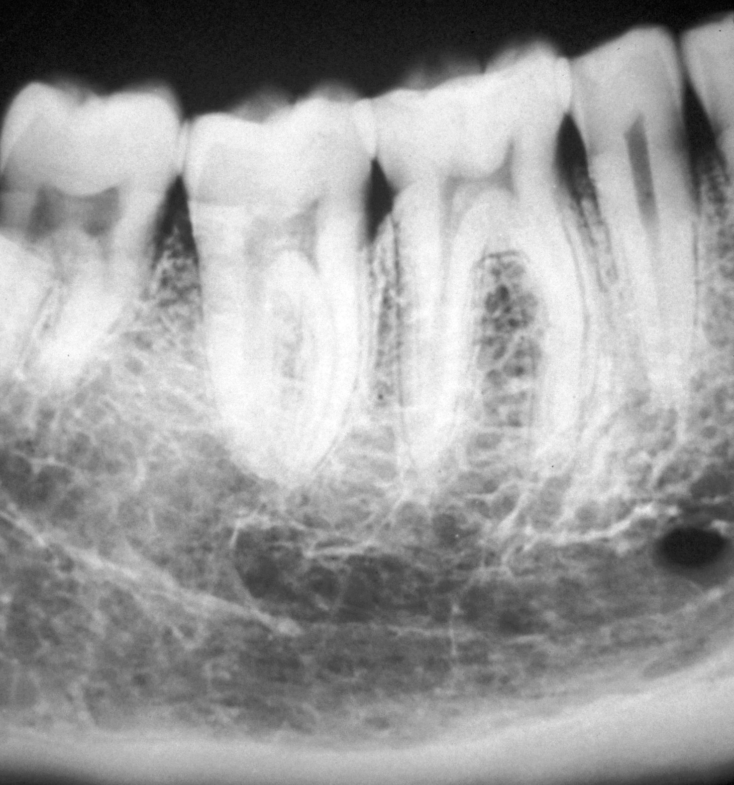
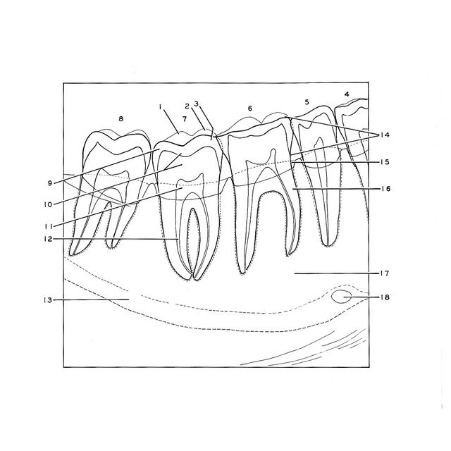
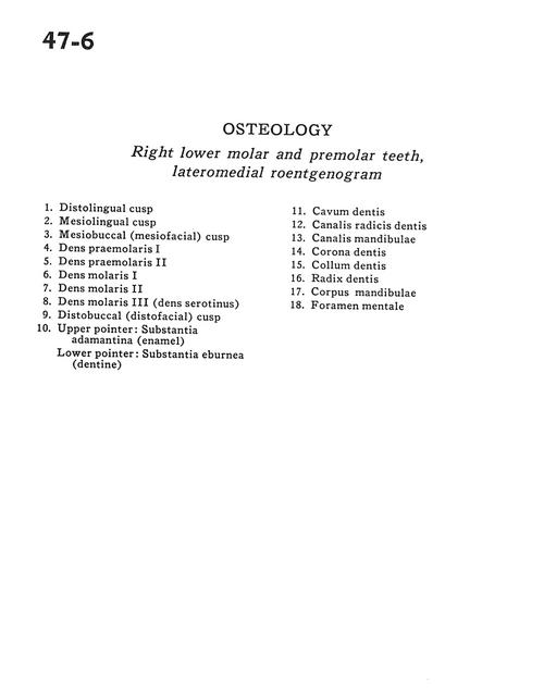
Osteology
Right lower molar and premolar teeth, lateromedial roentgenogram
Stanford holds the copyright to the David L. Bassett anatomical images and has assigned
Creative Commons license Attribution-Share
Alike 4.0 International to all of the images.
For additional information regarding use and permissions,
please contact the Medical History Center.
Image #47-6
Osteology
Right lower molar and premolar teeth, lateromedial roentgenogram
- Distolingual cusp
- Mesiolingual cusp
- Mesiobuccal (mesiofacial) cusp
- Premolar I
- Premolar II
- Molar I
- Molar II
- Molar III (dens serotinus)
- Distobuccal (distofacial) cusp
- Upper pointer: Substantia adamantina (enamel) Lower pointer: Substantia eburnea (dentine)
- Cavity
- Root canal
- Mandibular canal
- Crown
- Neck
- Root
- Body of mandible
- Mental foramen