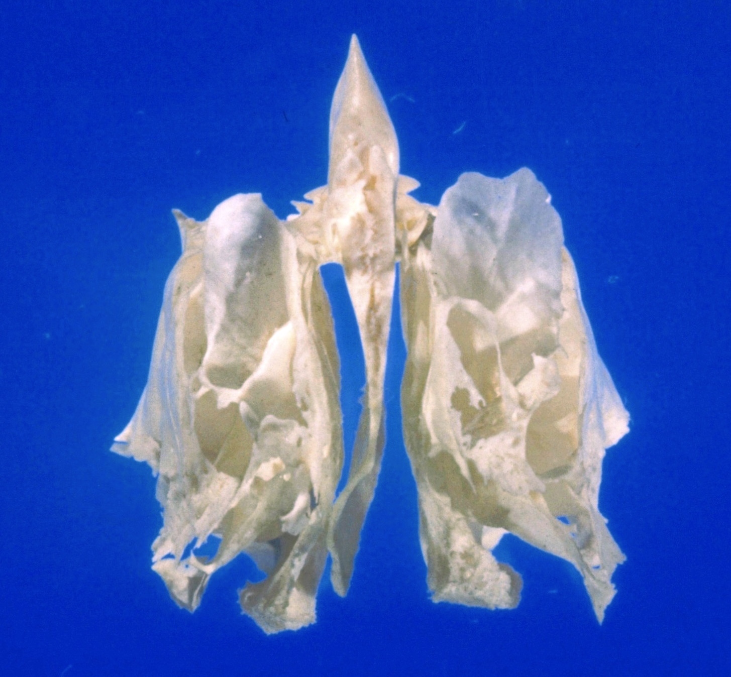
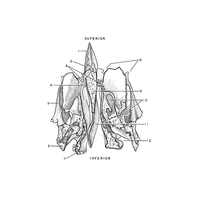
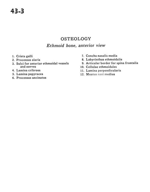
Osteology
Ethmoid bone, anterior view
Stanford holds the copyright to the David L. Bassett anatomical images and has assigned
Creative Commons license Attribution-Share
Alike 4.0 International to all of the images.
For additional information regarding use and permissions,
please contact the Medical History Center.
Image #43-3
Osteology
Ethmoid bone, anterior view
- Crista galli
- Alar process
- Sulci for anterior ethmoidal vessels and nerves
- Cribriform plate
- Lamina papyracea
- Uncinate process
- Middle nasal concha
- Ethmoidal labyrinth
- Articular border for frontal spine
- Ethmoidal cells
- Perpendicular plate
- Middle nasal meatus