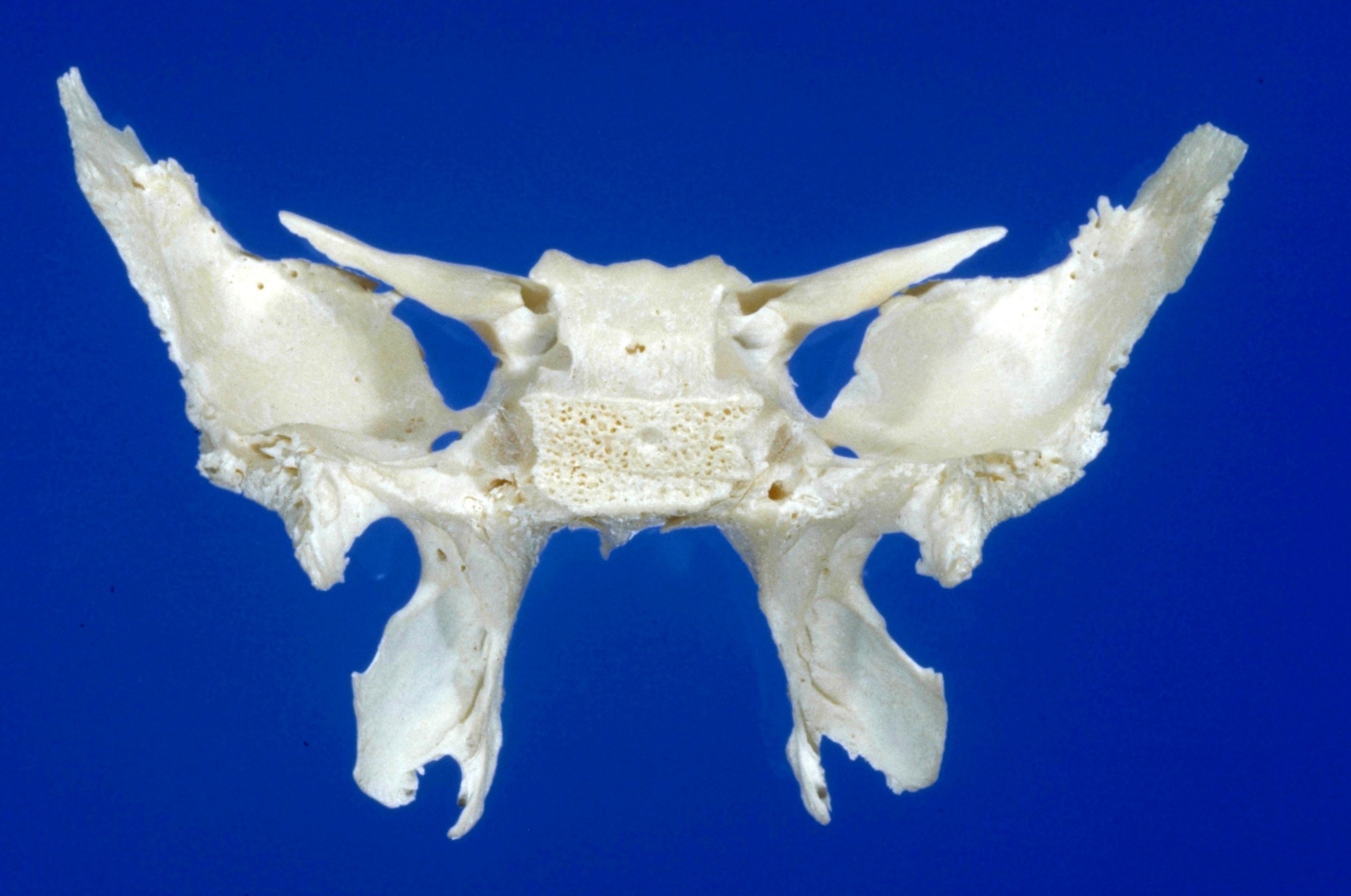
Osteology
Sphenoid bone, posterior view
Stanford holds the copyright to the David L. Bassett anatomical images and has assigned
Creative Commons license Attribution-Share
Alike 4.0 International to all of the images.
For additional information regarding use and permissions,
please contact the Medical History Center.
Image #42-4
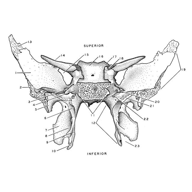
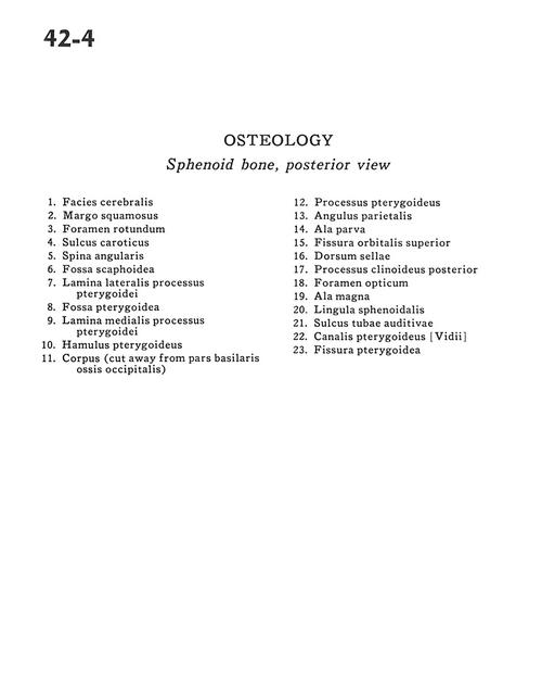
Osteology
Sphenoid bone, posterior view
- Cerebral surface
- Squamous margin
- Foramen rotundum
- Carotid sulcus
- Angular spine
- Scaphoid fossa
- Lateral plate of pterygoid process
- Pterygoid fossa
- Medial plate of pterygoid process
- Pterygoid hamulus
- Body (cut away from basal part occipital bone)
- Pterygoid process
- Parietal angle
- Lesser wing
- Superior orbital fissure
- Dorsum sellae
- Posterior clinoid process
- Optic foramen
- Greater wing
- Lingula
- Sulcus of auditory tube
- Pterygoid canal
- Pterygoid fissure