
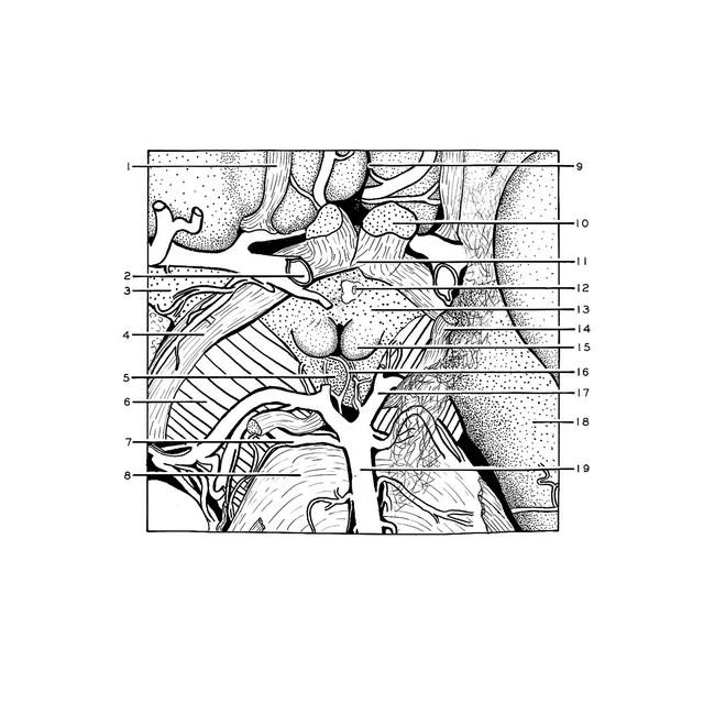
Exploration of the brain from its basal aspect
Arteries of interpeduncular fossa
Stanford holds the copyright to the David L. Bassett anatomical images and has assigned
Creative Commons license Attribution-Share
Alike 4.0 International to all of the images.
For additional information regarding use and permissions,
please contact the Medical History Center.
Image #4-6
Exploration of the brain from its basal aspect
Arteries of interpeduncular fossa
The numerous branches of the posterior cerebral arteries are illustrated in this close-up view as they enter the posterior perforated substance in the depths of the interpeduncular fossa. A piece of arachnoid membrane covers the left third nerve and cerebral peduncle (right side of view). The space seen between this membrane and the brain represents a portion of the interpeduncular cistern.
- Olfactory tract right
- Internal carotid artery
- Anterior perforated substance
- Optic tract
- Interpeduncular fossa
- Cerebral peduncle
- Superior cerebellar artery
- Pons
- Longitudinal fissure (cerebral)
- Optic nerve (II)
- Optic chiasm
- Infundibulum
- Tuber cinereum
- Oculomotor nerve (III)
- Mamillary body
- Medial central branch of posterior cerebral artery
- Posterior cerebral artery
- Hippocampal gyrus
- Basilar artery