Osteology
Hard palate, inferior surface
Stanford holds the copyright to the David L. Bassett anatomical images and has assigned
Creative Commons license Attribution-Share
Alike 4.0 International to all of the images.
For additional information regarding use and permissions,
please contact the Medical History Center.
Image #38-3
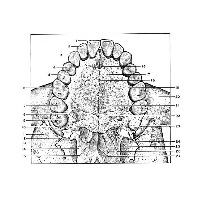
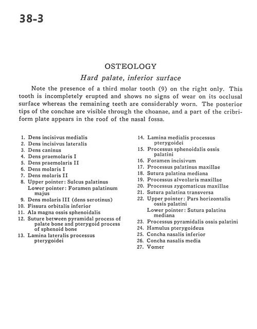
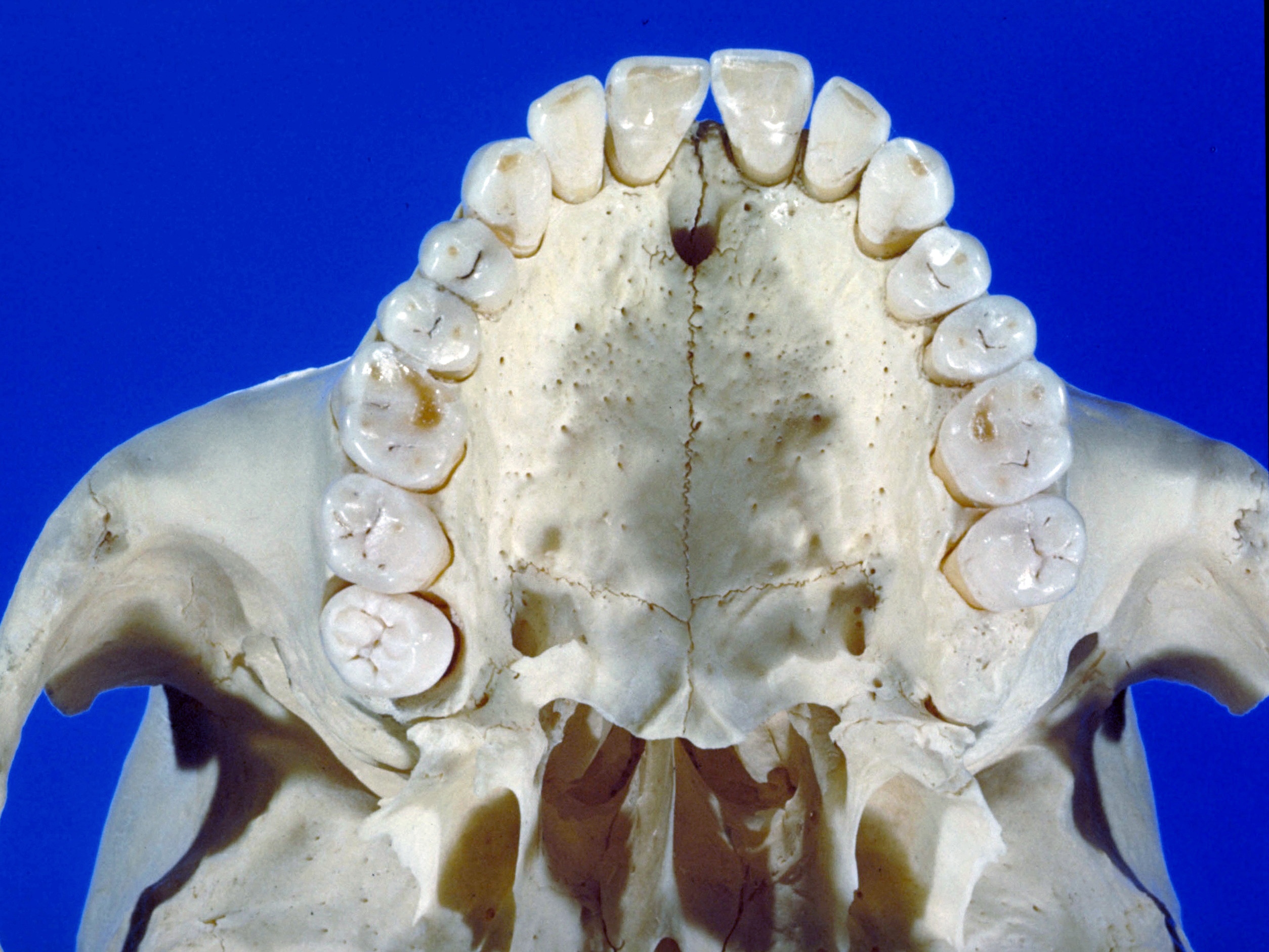
Osteology
Hard palate, inferior surface
Note the presence of a third molar tooth (9) on the right only. This tooth is incompletely erupted and shows no signs of wear on its occlusal surface whereas the remaining teet are considerably worn. The posterior tips of the conchae are visible through the choanae, and a part of the cribriform plate appears in the roof of the nasal fossa.
- Medial incisor
- Lateral incisor
- Canine
- First premolar
- Second pre-molar
- First molar
- Second molar
- Upper pointer: Palatine sulcus Lower pointer: Greater palatine foramen
- Third molar (serotinus)
- Inferior orbital fissure
- Greater wing of sphenoid
- Suture between pyramidal process of palate bone and pterygoid process of sphenoid bone
- Lateral plate of pterygoid process
- Medial plate of pterygoid process
- Sphenoid process palatine bone
- Incisive foramen
- Palatine process of maxilla
- Median palatine suture
- Alveolar process of maxilla
- Zygomatic process of maxilla
- Transverse palatine suture
- Upper pointer: Horizontal plate palatine bone Lower pointer: Median palatine suture
- Pyramidal process palatine bone
- Pterygoid hamulus
- Inferior nasal concha
- Middle nasal concha
- Vomer