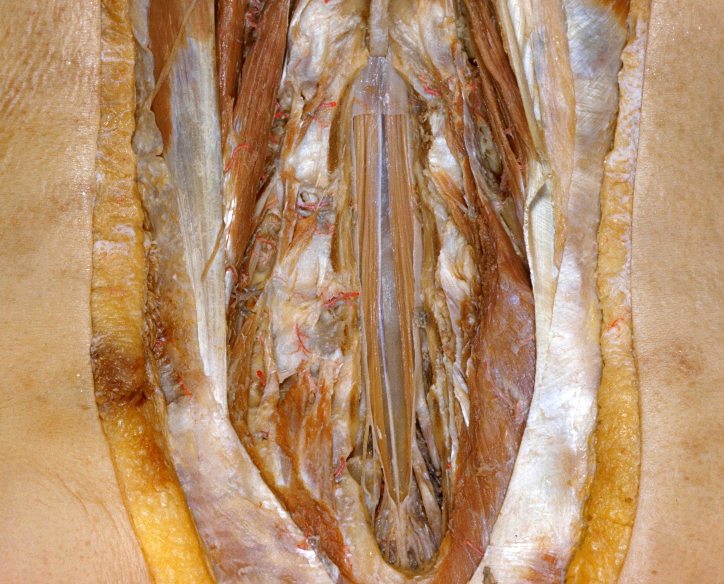
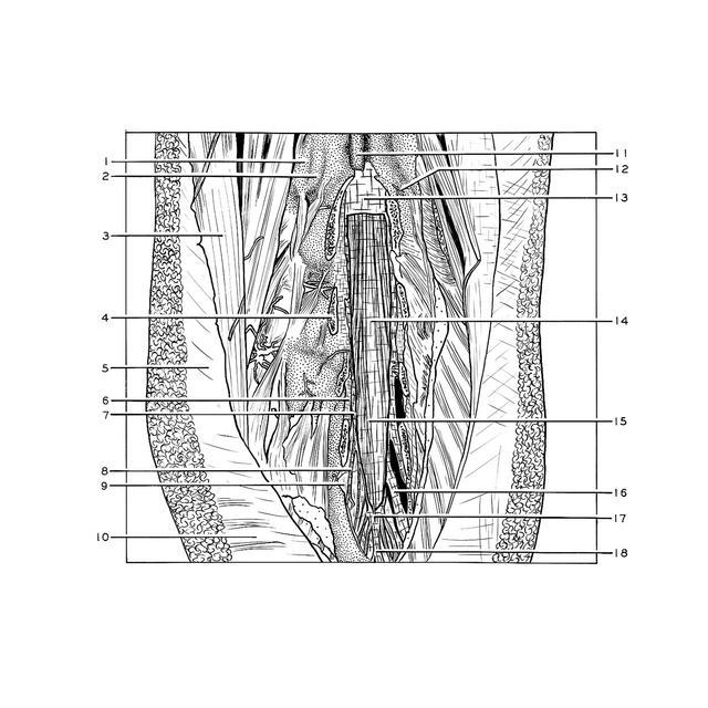
Exploration of the spinal cord and meninges in situ
Arachnoid membrane covering cauda equina
Stanford holds the copyright to the David L. Bassett anatomical images and has assigned
Creative Commons license Attribution-Share
Alike 4.0 International to all of the images.
For additional information regarding use and permissions,
please contact the Medical History Center.
Image #33-6
Exploration of the spinal cord and meninges in situ
Arachnoid membrane covering cauda equina
The dura has been cut away in such a manner that the arachnoid membrane remains intact. The filum terminale (15) appears as a white strand deep to the arachnoid. An extensive subarachnoid space surrounds nerve roots which form the cauda equina.
- Mamillary process of lumbar vertebra III
- Inferior articular process lumbar vertebra II
- Dorsal iliocostal muscle
- Arch of lumbar vertebra IV
- Lumbodorsal fascia
- Ligamentum flavum
- Epidural space and cut margin of dura
- Fibrous attachment of dura to sacrum
- Cystic enlargement of dura covering roots of second sacral nerve (note resorption of neighboring bone)
- Gluteus maximus muscle
- Spinous process lumbar vertebra II
- Branch lumbar artery
- Dura mater
- Arachnoid
- Filum terminale (beneath arachnoid)
- Root sacral nerve II
- Level at which spinal subarachnoid space terminates
- Filum spinal dura mater