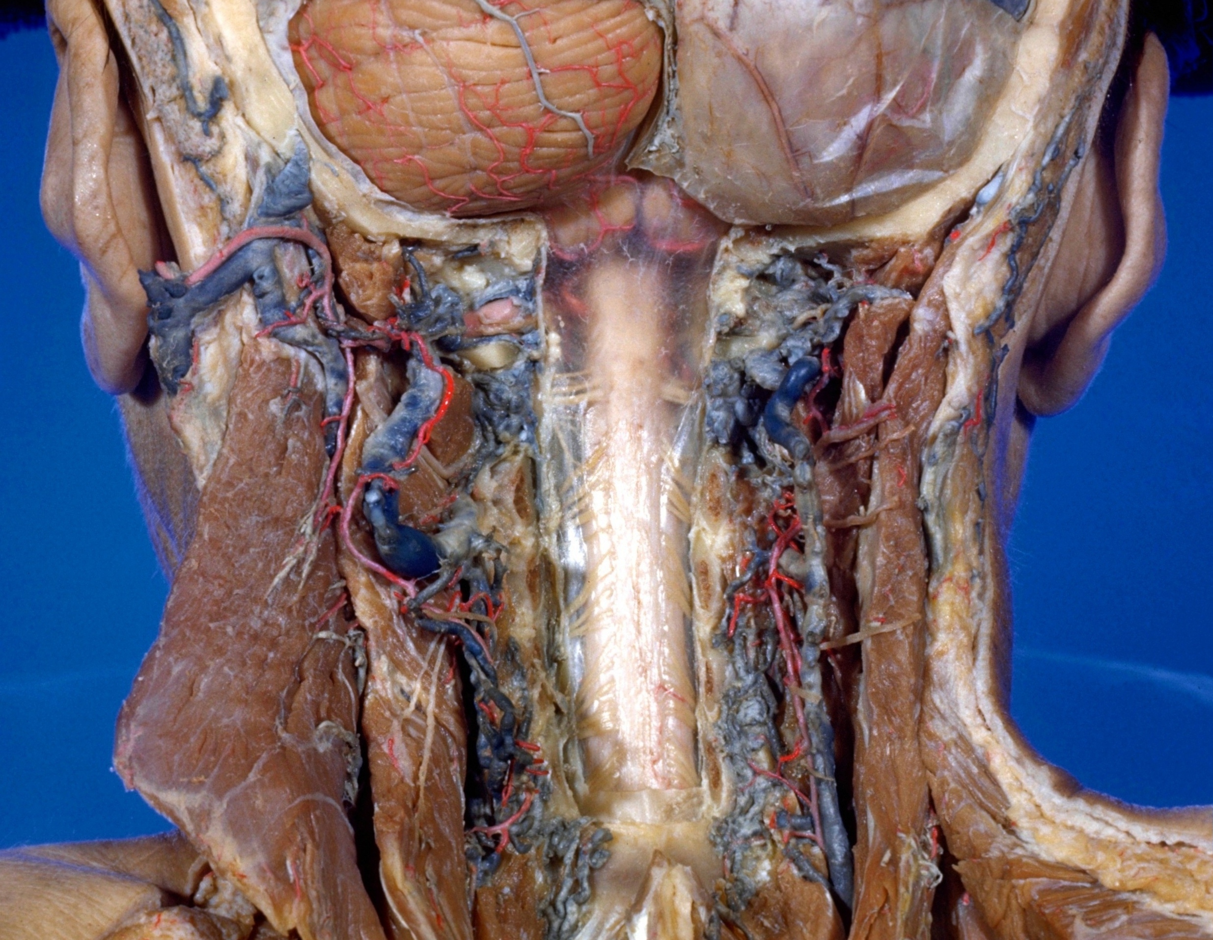
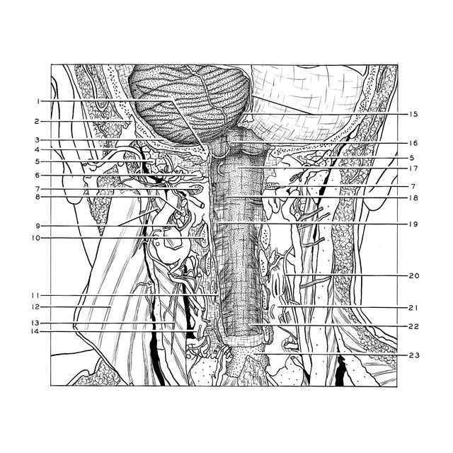
Exploration of the spinal cord and meninges in situ
Arachnoid in cervical region; cisterna magna
Stanford holds the copyright to the David L. Bassett anatomical images and has assigned
Creative Commons license Attribution-Share
Alike 4.0 International to all of the images.
For additional information regarding use and permissions,
please contact the Medical History Center.
Image #32-5
Exploration of the spinal cord and meninges in situ
Arachnoid in cervical region; cisterna magna
The dura mater has been cut away so that the transparent arachnoid membrane is exposed. The subarachnoid space increases in volume above the level of the second cervical dorsal roots. This is part of the cerebellomedullary cistern (cisterna magna).
- Occipital bone (pointer at margin of foramen magnum)
- Mastoid emissary
- Dura mater
- Occipital artery
- Superior obliquus capitis muscle
- Vertebral artery left
- Posterior arch of atlas (cut across)
- Inferior obliquus capitis muscle
- Greater occipital nerve
- Cervical vertebral arches II (cut across)
- Dura mater
- Splenius capitis muscle
- Motor nerve to semispinalis capitis muscle
- Posterior vertebral venous plexus
- Occipital sinus and falx cerebelli
- Arachnoid covering cerebellar tonsil
- Cerebellomedullary cistern
- Dorsal root cervical nerve II
- Accessory nerve (spinal root)
- Deep cervical artery
- Posterior vertebral venous plexus
- Dorsal root cervical nerve VI
- Arch of cervical vertebra VII