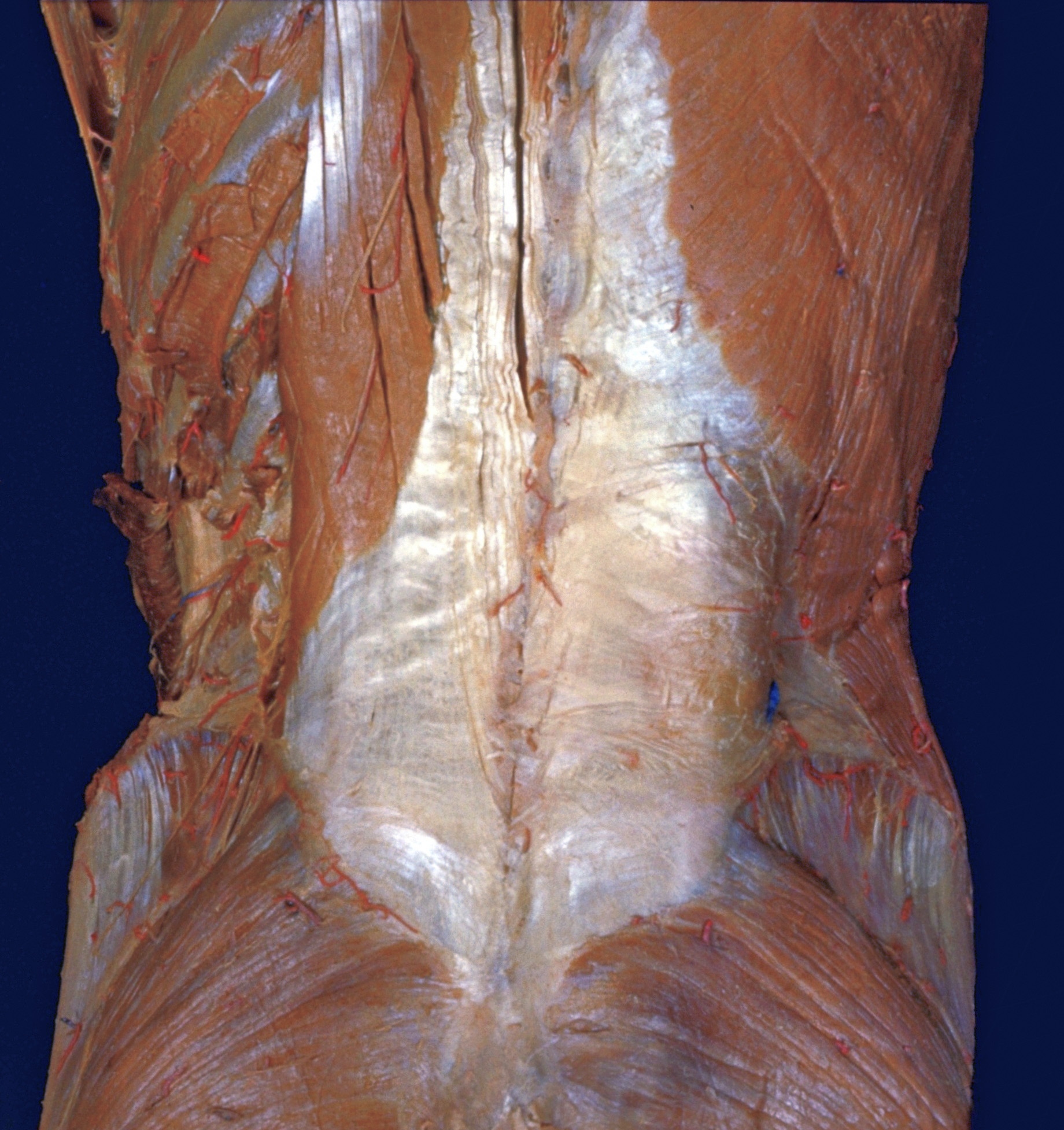
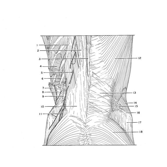
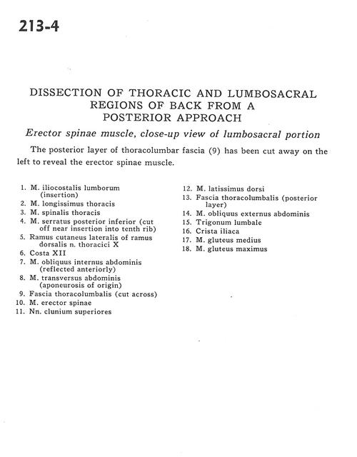
Dissection of thoracic and lumbosacral regions of back from a posterior approach
Erector spinae muscle, close-up view of lumbosacral portion
Stanford holds the copyright to the David L. Bassett anatomical images and has assigned
Creative Commons license Attribution-Share
Alike 4.0 International to all of the images.
For additional information regarding use and permissions,
please contact the Medical History Center.
Image #213-4
Dissection of thoracic and lumbosacral regions of back from a posterior approach
Erector spinae muscle, close-up view of lumbosacral portion
The posterior layer of thoracolumbar fascia (9)has been cut away on the left to reveal the erector spinae muscle.
- Iliocostalis lumborum muscle (insertion)
- Longissimus thoracis muscle
- Spinalis thoracis muscle
- Serratus posterior inferior muscle (cut off near insertion into tenth rib)
- Lateral cutaneous branch of dorsal branch thoracic nerve X
- Rib XII
- Internal oblique muscle (reflected anteriorly)
- Transverse abdominis muscle (aponeurosis of origin)
- Thoracolumbar fascia (cut across)
- Erector spinae muscle
- Superior cluneal nerves
- Latissimus dorsi muscle
- Thoracolumbar fascia (posterior layer)
- External oblique muscle
- Lumbar triangle (Petit's)
- Iliac crest
- Gluteus medius muscle
- Gluteus maximus muscle