Radiography
Radiograph of neck, left lateral view
Stanford holds the copyright to the David L. Bassett anatomical images and has assigned
Creative Commons license Attribution-Share
Alike 4.0 International to all of the images.
For additional information regarding use and permissions,
please contact the Medical History Center.
Image #210-2
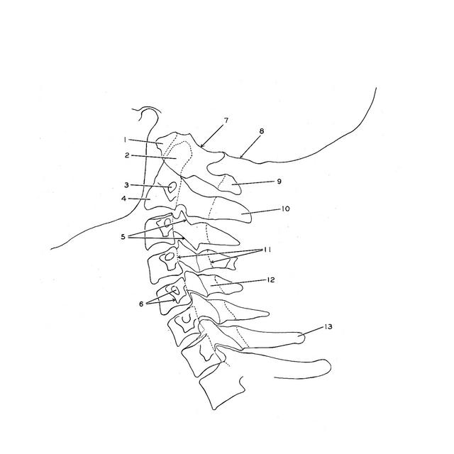
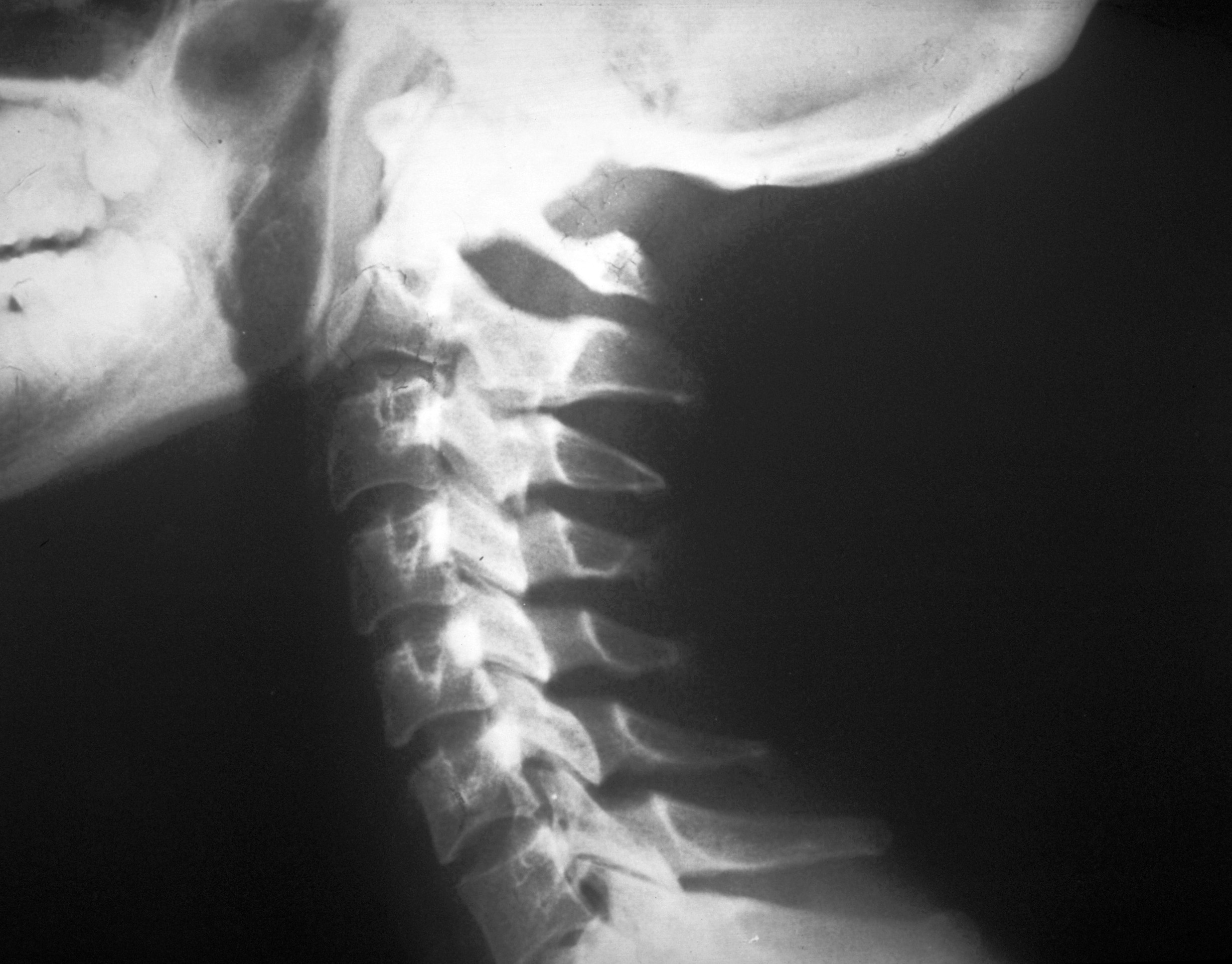
Radiography
Radiograph of neck, left lateral view
This film was obtained through the courtesy of Dr. Melvin J. Figley.
- Anterior arch of atlas
- Dens (axis)
- Transverse foramen axis
- Body of axis
- Upper pointer: Superior articular process vertebra C. III Lower pointer: Inferior articular process vertebra C. III
- Upper pointer: Transverse foramen vertebra C. V Lower pointer: Transverse process vertebra C. V
- Superior articular facet of atlas
- Occipital bone
- Posterior arch of atlas
- Spinous process axis
- Vertebral foramen (pointers indicate anterior and posterior borders of foramen of vertebra C. IV, outline of vertebral canal visible above and below this level)
- Lamina (arch of vertebra) C. V
- Spinous process vertebra C. VII (vertebra prominens)