Dissection of plantar aspect of left foot
Adductor hallucis muscle, general view
Stanford holds the copyright to the David L. Bassett anatomical images and has assigned
Creative Commons license Attribution-Share
Alike 4.0 International to all of the images.
For additional information regarding use and permissions,
please contact the Medical History Center.
Image #201-7
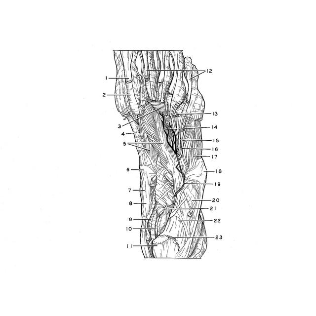
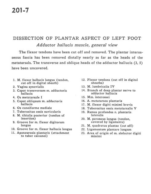
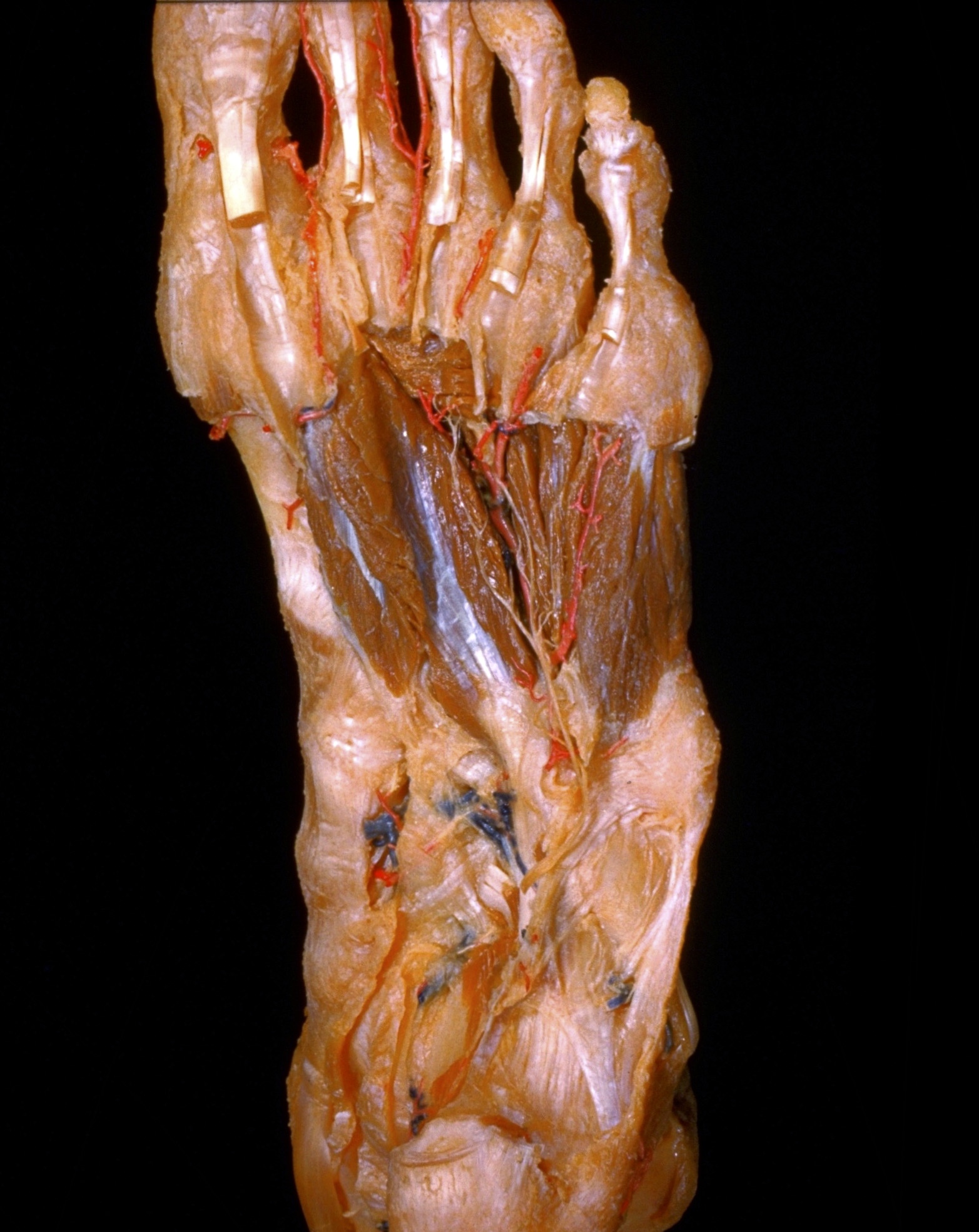
Dissection of plantar aspect of left foot
Adductor hallucis muscle, general view
The flexor tendons have been cut off and removed. The plantar interosseous fascia has been removed distally nearly as far as the heads of the metatarsals. The transverse and oblique heads of the adductor hallucis (3, 5) have been uncovered.
- Flexor hallucis longus muscle (tendon, cut off in digital sheath)
- Synovial sheath
- Transverse head of adductor hallucis muscle
- Metatarsal bone
- Oblique head of adductor hallucis muscle
- Medial cuneiform bone
- Tuberosity of navicular bone
- Tibialis posterior muscle (tendon of insertion)
- Groove for flexor digitorum longus muscle
- Groove for flexor hallucis longus muscle
- Plantar aponeurosis (attachment to tuberosity of calcaneus)
- Flexor tendons (cut off in digital sheaths)
- 4th lumbrical muscle
- Branch of deep plantar nerve to adductor hallucis
- Interosseous muscle
- Plantar metatarsal artery
- Flexor digiti minimi brevis muscle
- Tuberosity of 5th metatarsal bone
- Deep branch lateral plantar nerve
- Peroneus longus muscle (tendon, covered by ligaments)
- Quadratus plantae muscle (cut off)
- Long plantar ligament
- Area of origin of abductor digiti minimi muscle