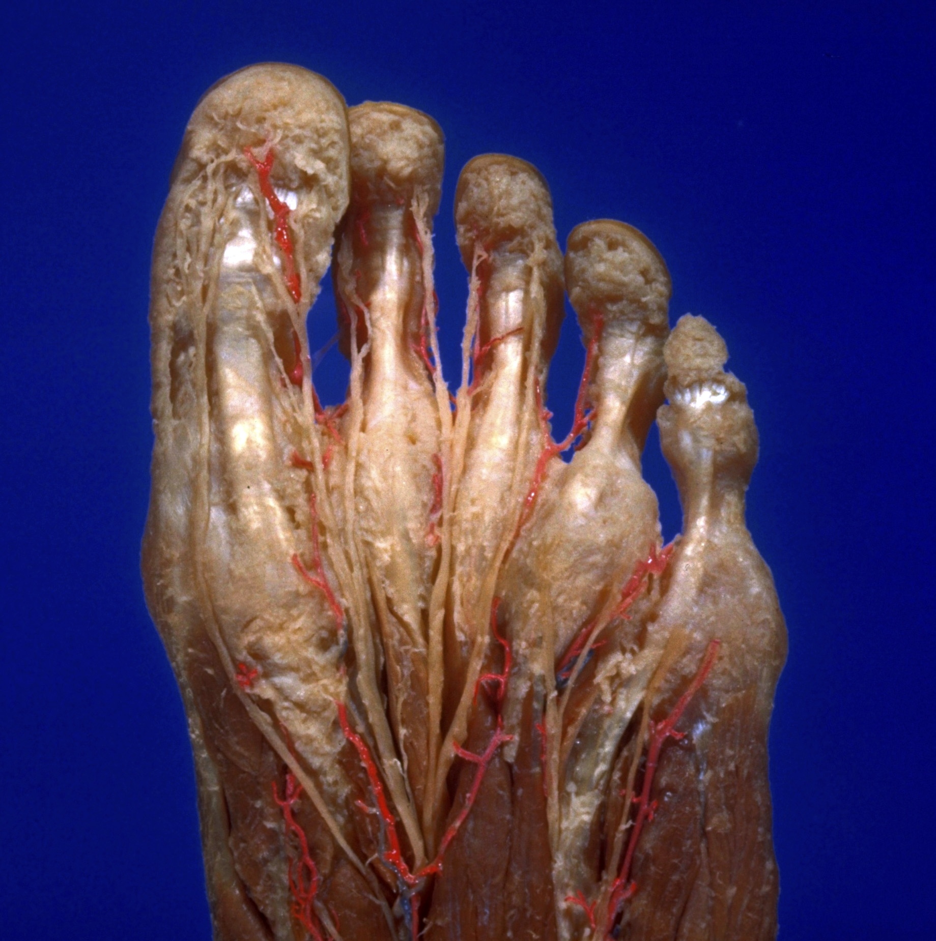
Dissection of plantar aspect of left foot
Digital tendon sheaths in relation to digital arteries and nerves, close-up view
Stanford holds the copyright to the David L. Bassett anatomical images and has assigned
Creative Commons license Attribution-Share
Alike 4.0 International to all of the images.
For additional information regarding use and permissions,
please contact the Medical History Center.
Image #200-4
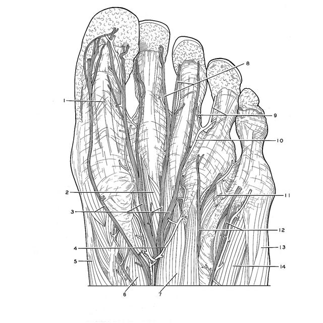
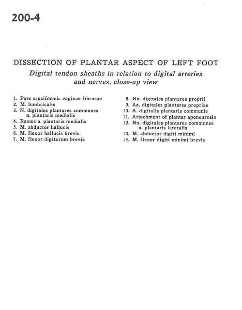
Dissection of plantar aspect of left foot
Digital tendon sheaths in relation to digital arteries and nerves, close-up view
- Fibrous sheath of pars cruciformis
- Lumbrical muscle
- Common plantar digital nerves and medial plantar nerve
- Branch of medial plantar artery
- Abductor hallucis muscle
- Flexor hallucis brevis muscle
- Flexor digitorum brevis muscle
- Proper plantar digital nerves
- Proper plantar digital arteries
- Common plantar digital artery
- Attachment of plantar aponeurosis
- Common plantar digital nerves and lateral plantar nerve
- Abductor digiti minimi muscle
- Flexor digiti minimi brevis muscle