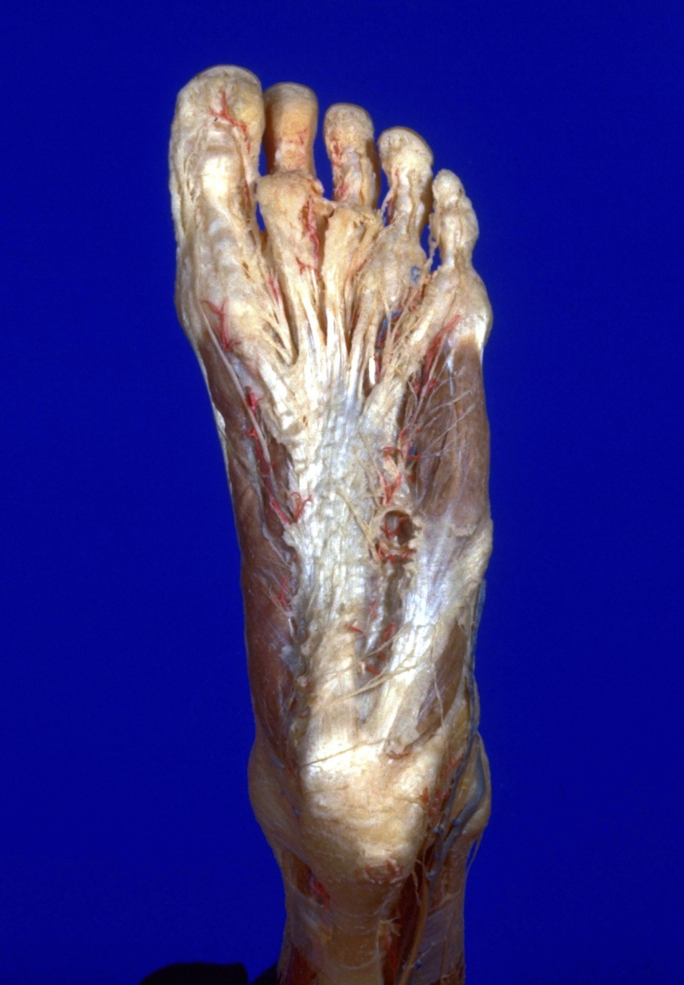
Dissection of plantar aspect of left foot
Plantar aponeurosis
Stanford holds the copyright to the David L. Bassett anatomical images and has assigned
Creative Commons license Attribution-Share
Alike 4.0 International to all of the images.
For additional information regarding use and permissions,
please contact the Medical History Center.
Image #200-1
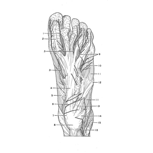
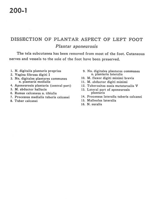
Dissection of plantar aspect of left foot
Plantar aponeurosis
The tela subcutanea has been removed from most of the foot. Cutaneous nerves and vessels to the sole of the foot have been preserved.
- Proper plantar digital nerve
- Fibrous sheath of 1st digit
- Common plantar digital nerves and medial plantar nerve
- Plantar aponeurosis (central part)
- Abductor hallucis muscle
- Calcaneal branch of tibial nerve
- Medial process of tuberosity of calcaneus
- Tuberosity of calcaneus
- Common plantar digital nerves and lateral plantar nerve
- Flexor digiti minimi brevis muscle
- Abductor digiti minimi muscle
- Tuberosity of 5th metatarsal bone
- Lateral part of plantar aponeurosis
- Lateral tubercle process of calcaneus bone
- Lateral malleolus
- Sural nerve