Exploration of those parts of the brain supplied by the posterior cerebral artery
Optic tract and lateral geniculate body
Stanford holds the copyright to the David L. Bassett anatomical images and has assigned
Creative Commons license Attribution-Share
Alike 4.0 International to all of the images.
For additional information regarding use and permissions,
please contact the Medical History Center.
Image #20-7
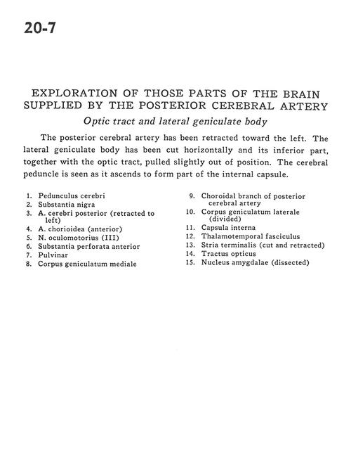
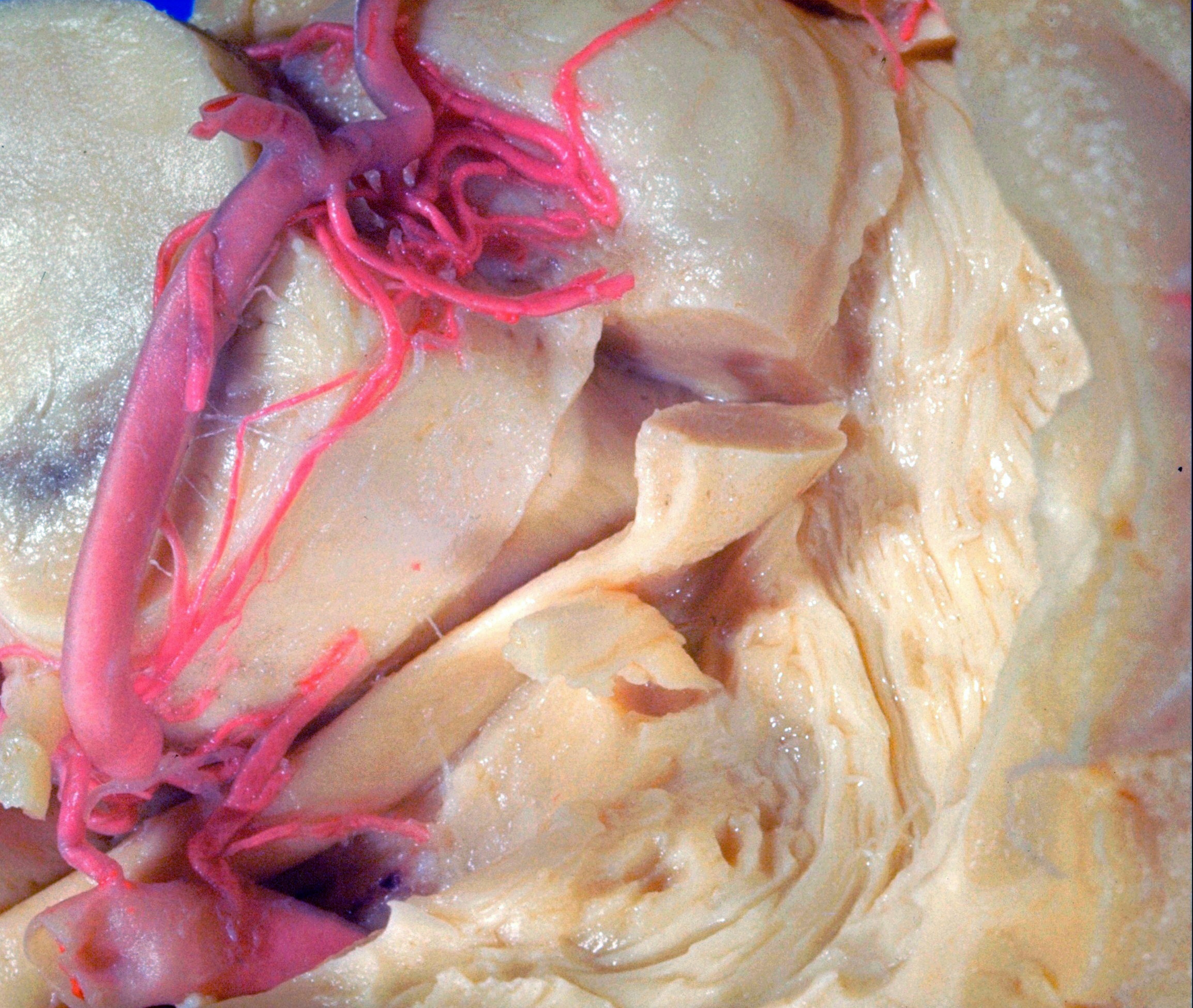
Exploration of those parts of the brain supplied by the posterior cerebral artery
Optic tract and lateral geniculate body
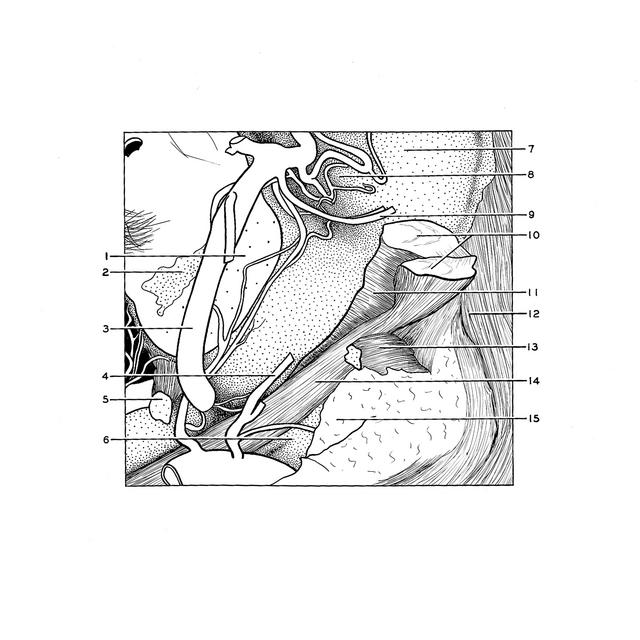
The posterior cerebral artery has been retracted toward the left. The lateral geniculate body has been cut horizontally and its inferior part, together with the optic tract, pulled slightly out of position. The cerebral peduncle is seen as it ascends to form part of the internal capsule.
- Cerebral peduncle
- Substantia nigra
- Posterior cerebral artery (retracted to the left)
- Choroidal artery (anterior)
- Oculomotor nerve (III)
- Anterior perforated substance
- Pulvinar
- Medial geniculate body
- Choroidal branch of posterior cerebral artery
- Lateral geniculate body (divided)
- Internal capsule
- Thalamotemporal fasciculus
- Stria terminalis (cut and retracted)
- Optic tract
- Amygdaloid nucleus (dissected)