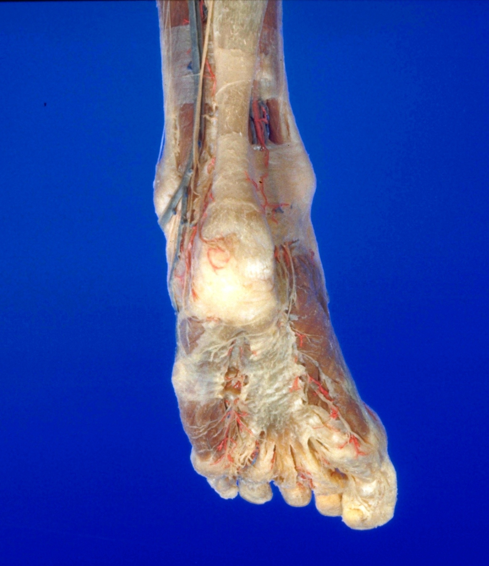
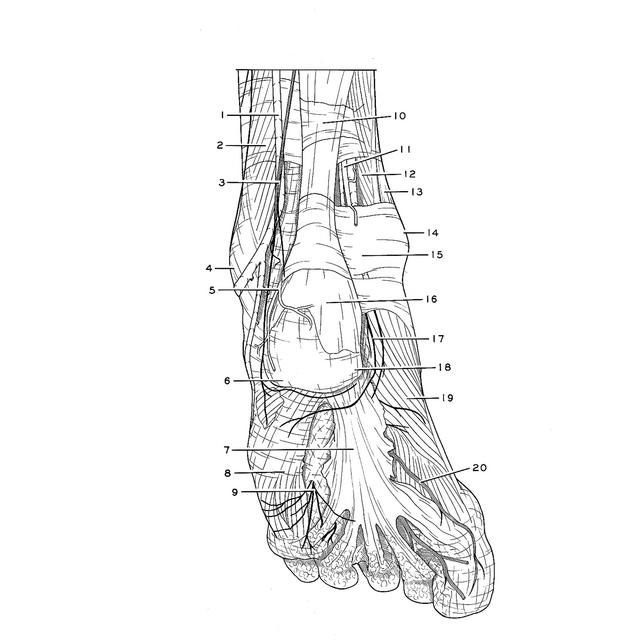
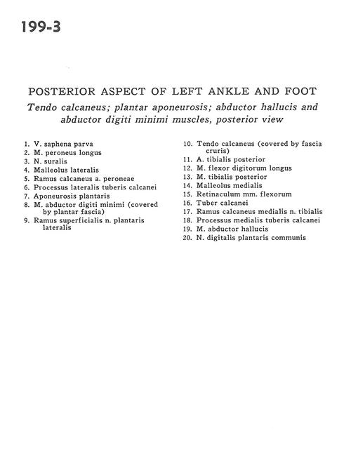
Posterior aspect of left ankle and foot
Tendo calcaneus; plantar aponeurosis; abductor hallucis and abductor digiti minimi muscles, posterior view
Stanford holds the copyright to the David L. Bassett anatomical images and has assigned
Creative Commons license Attribution-Share
Alike 4.0 International to all of the images.
For additional information regarding use and permissions,
please contact the Medical History Center.
Image #199-3
Posterior aspect of left ankle and foot
Tendo calcaneus; plantar aponeurosis; abductor hallucis and abductor digiti minimi muscles, posterior view
- Lesser saphenous vein
- Peroneus longus muscle
- Sural nerve
- Lateral malleolus
- Calcaneal branch of peroneal artery
- Lateral tubercle process of calcaneus bone
- Plantar aponeurosis
- Abductor digiti minimi muscle (covered by plantar fascia)
- Superficial branch of lateral plantar nerve
- Tendo calcaneus (covered by crural fascia)
- Posterior tibial artery
- Flexor digitorum longus muscle
- Tibialis posterior muscle
- Medial malleolus
- Flexor retinaculum
- Tuberosity of calcaneus
- Calcaneal branch of medial tibial nerve
- Medial process of tuberosity of calcaneus
- Abductor hallucis muscle
- Common plantar digital nerve