Dissection of dorsolateral aspect of left foot and ankle
Nerve supply to extensor digitorum brevis and extensor hallucis brevis muscles
Stanford holds the copyright to the David L. Bassett anatomical images and has assigned
Creative Commons license Attribution-Share
Alike 4.0 International to all of the images.
For additional information regarding use and permissions,
please contact the Medical History Center.
Image #198-1
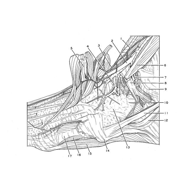
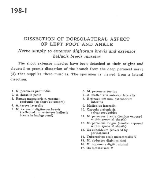
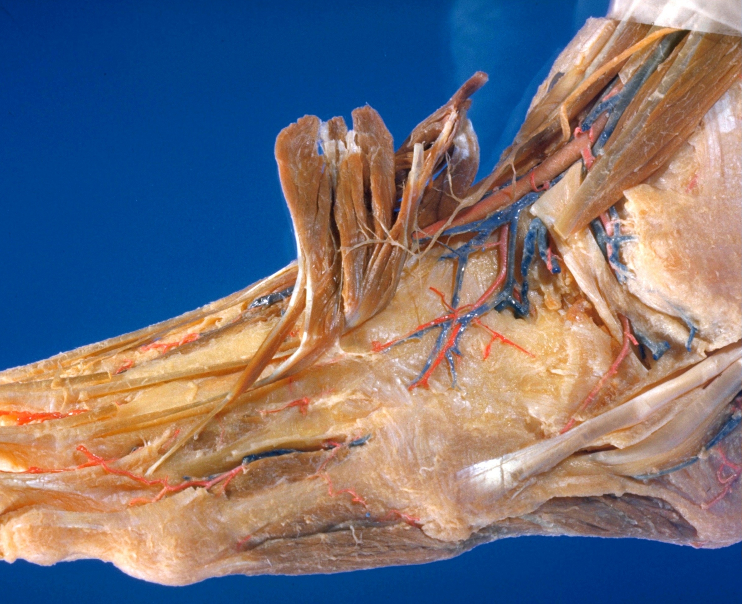
Dissection of dorsolateral aspect of left foot and ankle
Nerve supply to extensor digitorum brevis and extensor hallucis brevis muscles
The short extensor muscles have been detached at their origins and elevated to permit dissection of the branch from the deep peroneal nerve (3) that supplies these muscles. The specimen is viewed from a lateral direction.
- Deep peroneal nerve
- Dorsalis pedis artery
- Muscular branch of deep peroneal nerve (to short extensors)
- Lateral tarsal artery
- Extensor digitorum brevis muscle (reflected, extensor hallucis brevis muscle in background)
- Peroneus tertius muscle
- Anterior lateral malleolar artery
- Inferior extensor retinaculum
- Lateral malleolus
- Calcaneocuboid articular capsule
- Peroneus brevis muscle (tendon exposed within synovial sheath)
- Peroneus longus muscle (tendon exposed within synovial sheath)
- Cuboid bone (covered by periosteum)
- Tuberosity of 5th metatarsal bone
- Abductor digiti minimi muscle
- Opponens digiti minimi muscle
- 5th metatarsal bone