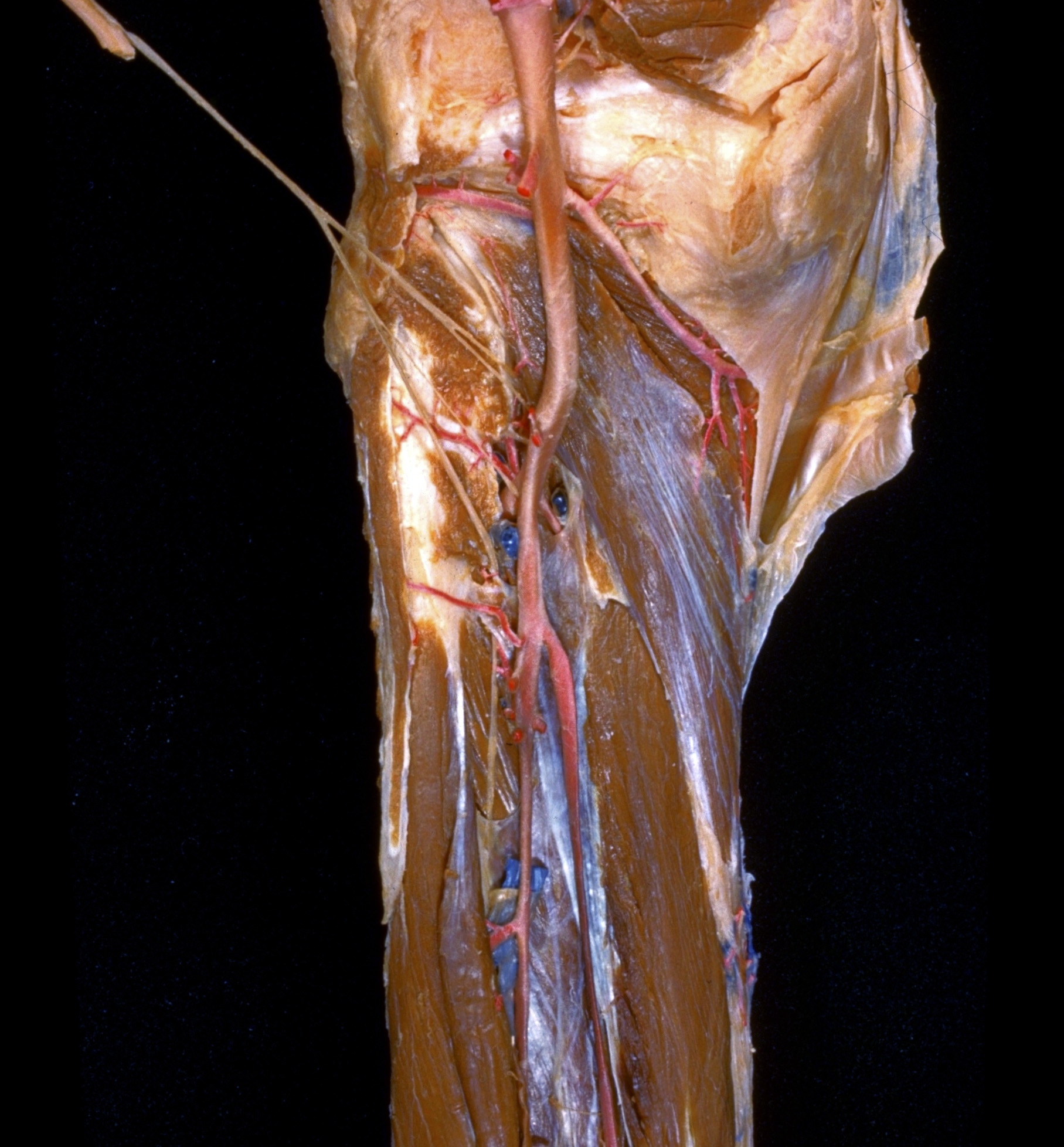
Dissection of posterior aspect of left leg
Popliteus muscle, close-up view
Stanford holds the copyright to the David L. Bassett anatomical images and has assigned
Creative Commons license Attribution-Share
Alike 4.0 International to all of the images.
For additional information regarding use and permissions,
please contact the Medical History Center.
Image #196-1
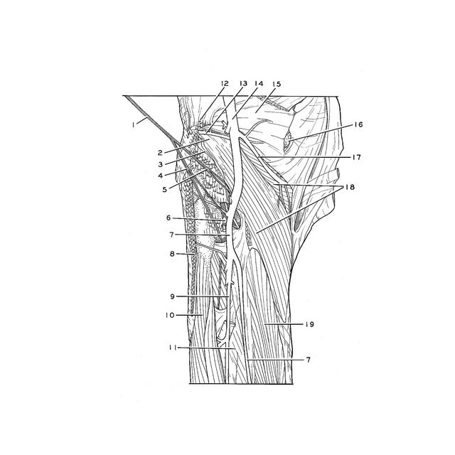
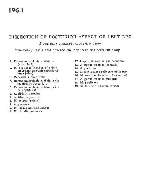
Dissection of posterior aspect of left leg
Popliteus muscle, close-up view
The heavy fascia that covered the popliteus has been cut away.
- Muscular branch of tibial nerve (stretched)
- Popliteus muscle (tendon of origin emerging through capsule of knee joint)
- Subpopliteal recess
- Muscular branch of tibial nerve (to tibialis posterior muscle)
- Muscular branch of tibial nerve (to popliteus muscle)
- Anterior tibial artery
- Posterior tibial artery
- Soleus muscle (origin)
- Peroneal artery
- Flexor hallucis longus muscle
- Tibialis posterior muscle
- Lateral head of gastrocnemius muscle
- Lateral inferior genicular artery
- Popliteal artery
- Oblique popliteal ligament
- Semimembranosus muscle (insertion)
- Medial inferior genicular artery
- Popliteus muscle
- Flexor digitorum longus muscle