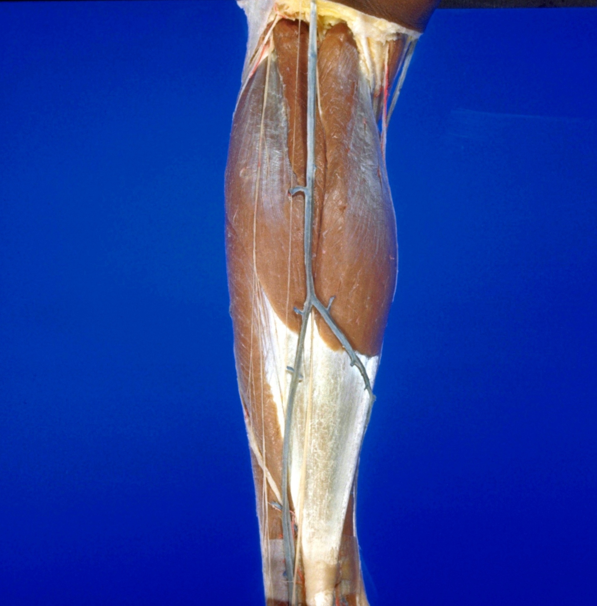
Dissection of posterior aspect of left leg
Muscles of leg, posterior view
Stanford holds the copyright to the David L. Bassett anatomical images and has assigned
Creative Commons license Attribution-Share
Alike 4.0 International to all of the images.
For additional information regarding use and permissions,
please contact the Medical History Center.
Image #194-4
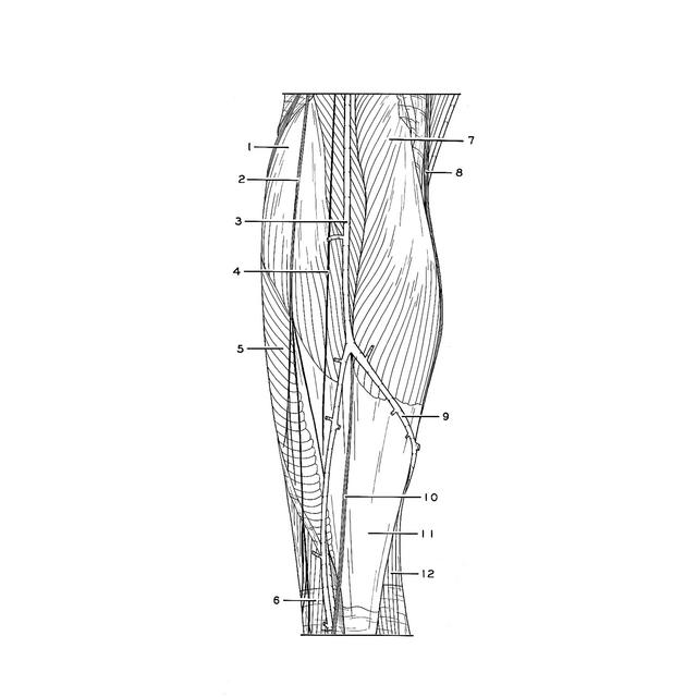
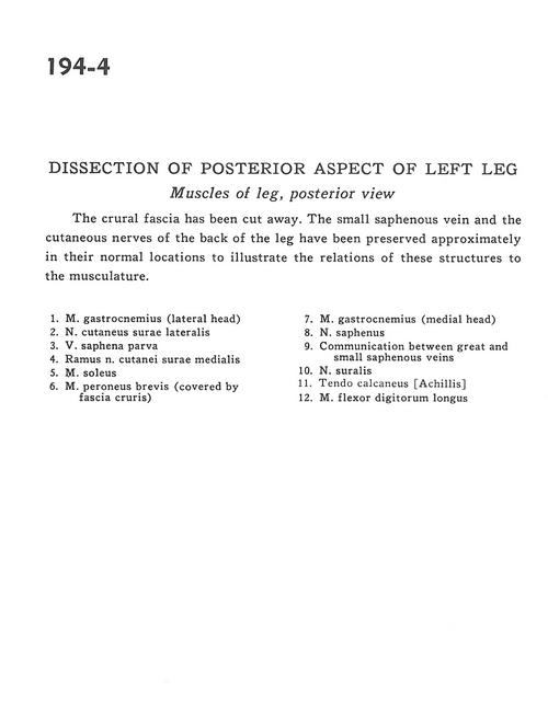
Dissection of posterior aspect of left leg
Muscles of leg, posterior view
The crural fascia has been cut away. The small saphenous vein and the cutaneous nerves of the back of the leg have been preserved approximately to their normal locations to illustrate the relations of these structures to the musculature.
- Gastrocnemius muscle (lateral head)
- Lateral cutaneous sural nerve
- Lesser saphenous vein
- Branch of medial sural cutaneous nerve
- Soleus muscle
- Peroneus brevis muscle (covered by crural fascia)
- Gastrocnemius muscle (medial head)
- Saphenous nerve
- Communication between great and small saphenous veins
- Sural nerve
- Tendo calcaneus (Achilles)
- Flexor digitorum longus muscle