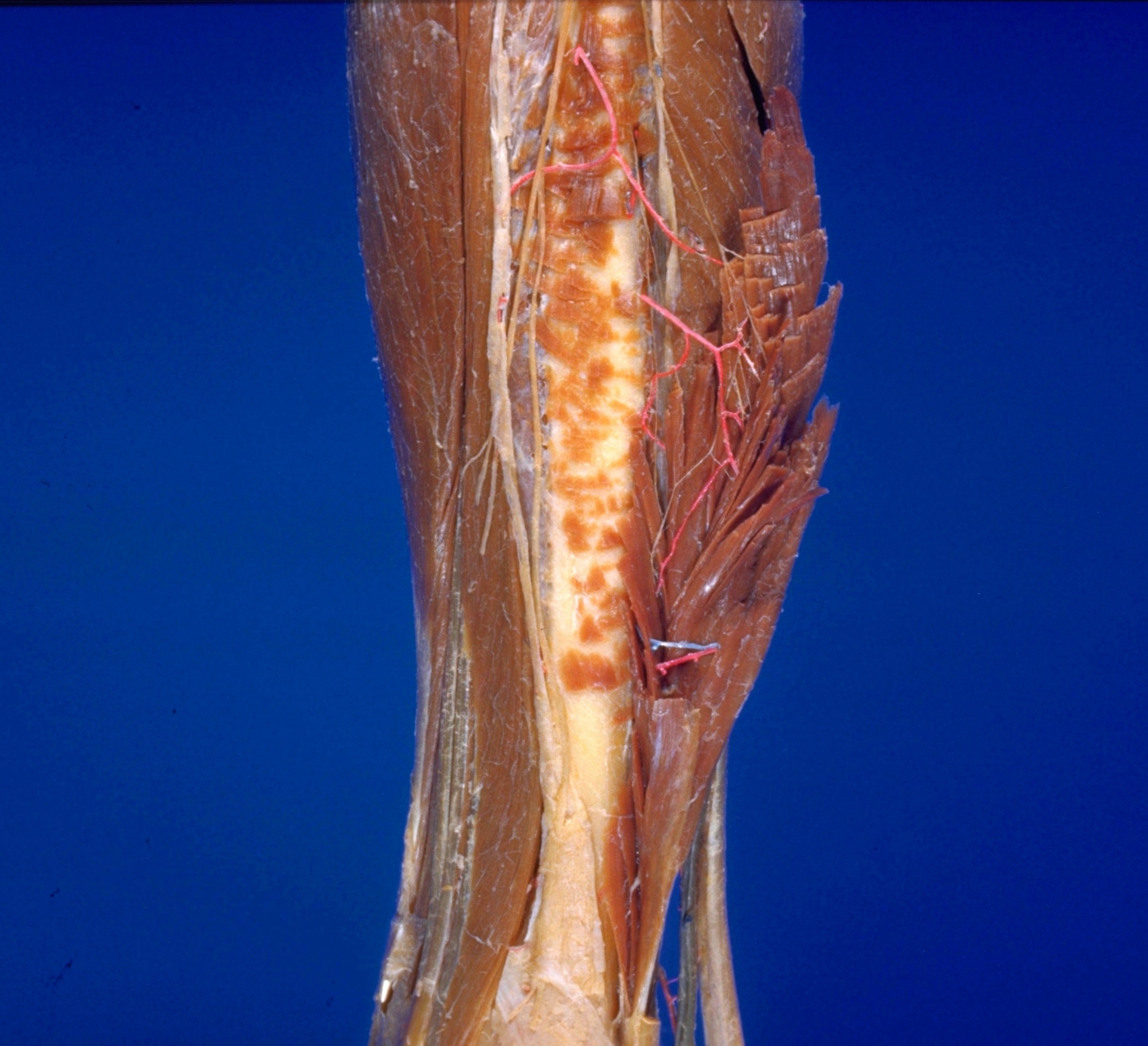
Dissection of lateral aspect of left leg
Nerves and blood vessels to peroneus brevis muscle
Stanford holds the copyright to the David L. Bassett anatomical images and has assigned
Creative Commons license Attribution-Share
Alike 4.0 International to all of the images.
For additional information regarding use and permissions,
please contact the Medical History Center.
Image #193-7
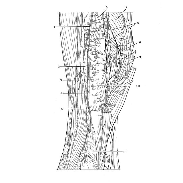
Dissection of lateral aspect of left leg
Nerves and blood vessels to peroneus brevis muscle
The peroneus brevis has been detached from its origins on the fibula (10) and anterior intermuscular septum (2). The muscle has been dissected and turned posteriorly to reveal the course of nerves and blood vessels into muscle substance.
- Superficial peroneal nerve
- Anterior intermuscular septum
- Dorsal medial cutaneous nerve
- Dorsal intermediate cutaneous nerve (this nerve passed along superficial surface of peroneus brevis)
- Peroneus tertius muscle
- Branch of anterior tibial artery
- Soleus muscle (in background)
- Muscular branch of superficial peroneal nerve (to peroneus brevis muscle)
- Peroneus brevis muscle (reflected)
- Lateral surface of fibula
- Subcutaneous surface of fibula