Dissection of lateral aspect of left leg
Peroneus brevis muscle
Stanford holds the copyright to the David L. Bassett anatomical images and has assigned
Creative Commons license Attribution-Share
Alike 4.0 International to all of the images.
For additional information regarding use and permissions,
please contact the Medical History Center.
Image #193-5
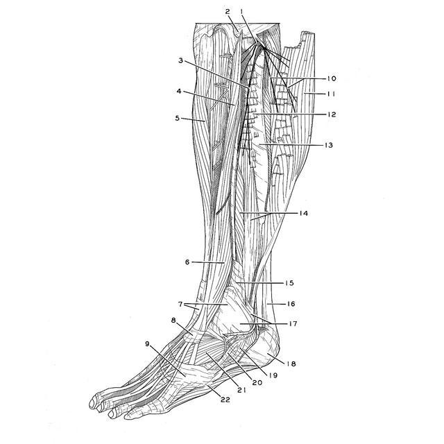
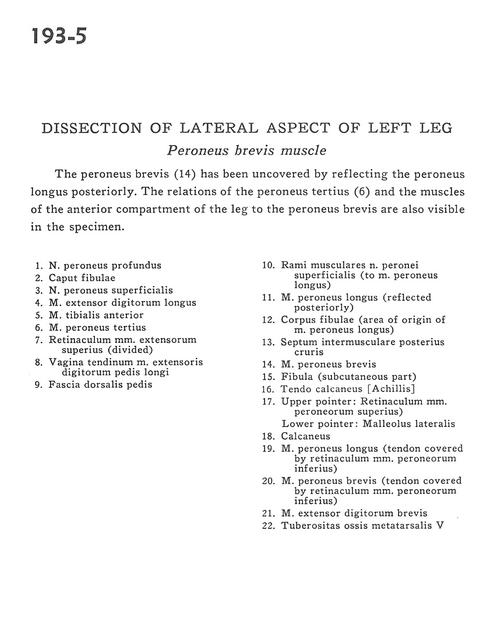
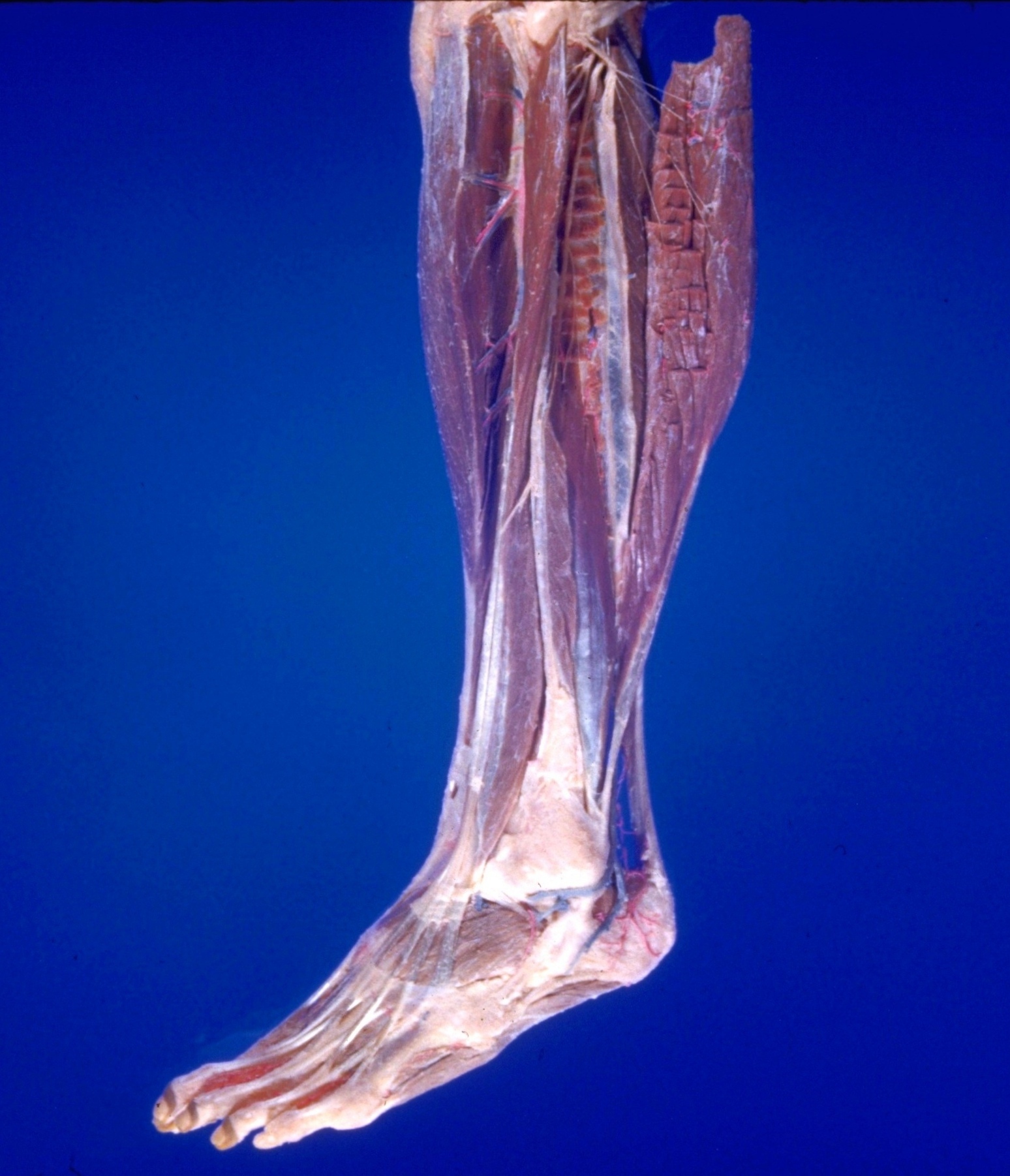
Dissection of lateral aspect of left leg
Peroneus brevis muscle
The peroneus brevis (14) has been uncovered by reflecting the peroneus longus posteriorly. The relations of the peroneus tertius (6) and the muscles of the anterior compartment of the leg to the peroneus brevis are also visible in the specimen.
- Deep peroneal nerve
- Head of fibula
- Superficial peroneal nerve
- Extensor digitorum longus muscle
- Tibialis anterior muscle
- Peroneus tertius muscle
- Superior extensor retinaculum (divided)
- Tendinous sheath of extensor digitorum pedis longus muscle
- Fascia of dorsalis pedis
- Muscular branch of superficial peroneal nerve (to peroneus longus muscle)
- Peroneus longus muscle (reflected posteriorly)
- Body of fibula (area of origin of peroneus longus muscle)
- Posterior intermuscular septum
- Peroneus brevis muscle
- Fibula (subcutaneous part)
- Tendo calcaneus (Achilles)
- Upper pointer: Superior peroneal retinaculum Lower pointer: Lateral malleolus
- Calcaneus
- Peroneus longus muscle (tendon covered by inferior peroneal retinaculum)
- Peroneus brevis muscle (tendon covered by inferior peroneal retinaculum)
- Extensor digitorum brevis muscle
- Tuberosity of 5th metatarsal bone