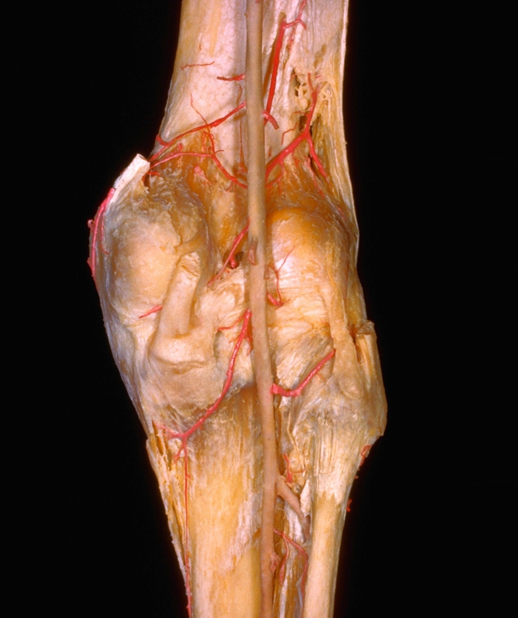
Dissection of knee
Capsule, ligaments and arteries of right knee, posterior view
Stanford holds the copyright to the David L. Bassett anatomical images and has assigned
Creative Commons license Attribution-Share
Alike 4.0 International to all of the images.
For additional information regarding use and permissions,
please contact the Medical History Center.
Image #190-2
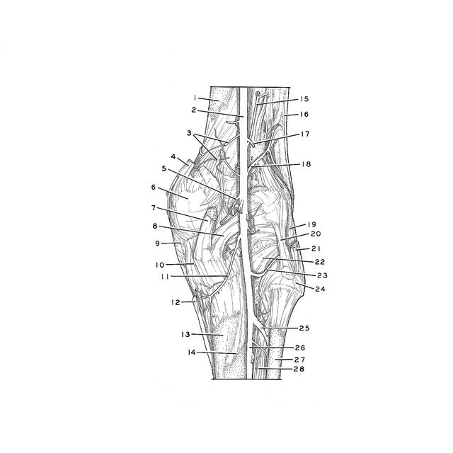
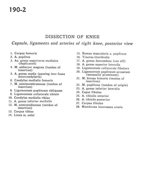
Dissection of knee
Capsule, ligaments and arteries of right knee, posterior view
- Body of femur
- Popliteal artery
- Lateral superior genicular arteries (duplicated)
- Adductor magnus muscle (tendon of insertion)
- Medial genicular artery (passing into intercondylar fossa)
- Medial condyle of femur
- Semimembranosus muscle (tendon of insertion)
- Oblique popliteal ligament
- Collateral ligament of tibia
- Medial condyle of tibia
- Lateral inferior genicular artery
- Semitendinosus muscle (tendon of insertion)
- Body of tibia
- Soleal line
- Muscular branch of popliteal artery
- Iliotibial tract
- Descending genicular artery (cut off)
- Medial superior genicular artery
- Collateral ligament of fibula
- Popliteal arcuate ligament
- Biceps fern oris muscle (tendon of insertion)
- Popliteus muscle (tendon of origin)
- Medial inferior genicular artery
- Head of fibula
- Anterior tibial artery
- Posterior tibial artery
- Body of fibula
- Interosseous membrane of leg