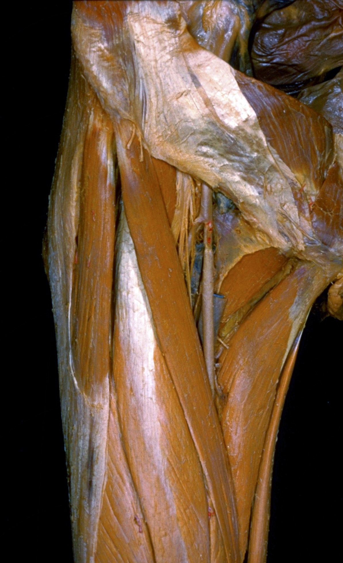
Dissection of anterior and medial aspects of thigh
Right tensor fasciae latae muscle, anterior view
Stanford holds the copyright to the David L. Bassett anatomical images and has assigned
Creative Commons license Attribution-Share
Alike 4.0 International to all of the images.
For additional information regarding use and permissions,
please contact the Medical History Center.
Image #185-7
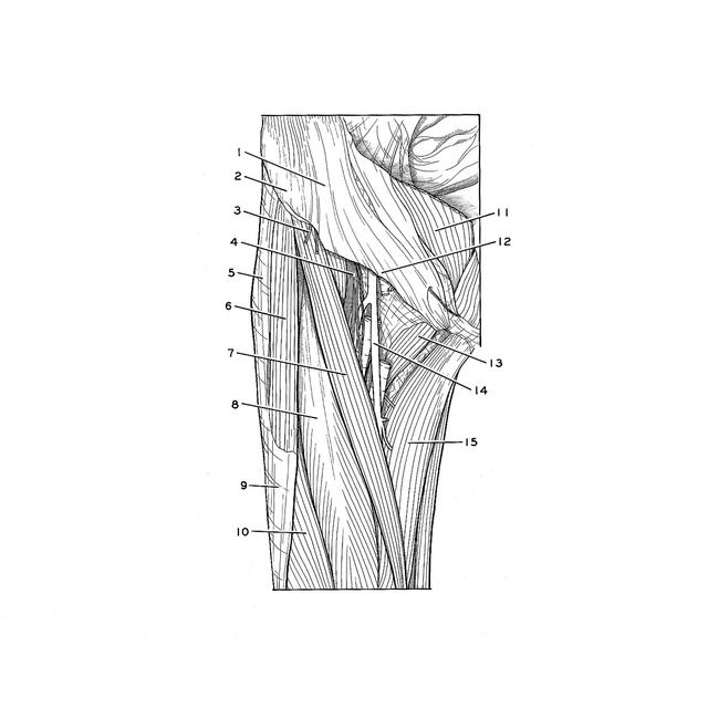
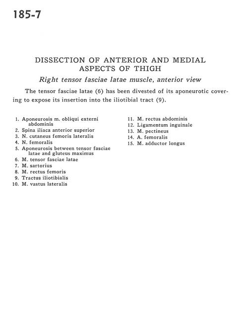
Dissection of anterior and medial aspects of thigh
Right tensor fasciae latae muscle, anterior view
The tensor fasciae latae (6) has been divested of its aponeurotic covering to expose its insertion into the iliotibial tract (9).
- Aponeurosis of External oblique muscle
- Anterior superior iliac spine
- Lateral femoral cutaneous nerve
- Femoral nerve
- Aponeurosis between tensor fasciae latae and gluteus maximus
- Tensor fasciae latae muscle
- Sartorius muscle
- Rectus femoris muscle
- Iliotibial tract
- Vastus lateralis muscle
- Rectus abdominis muscle
- Inguinal ligament
- Pectineus muscle
- Femoral artery
- Adductor longus muscle