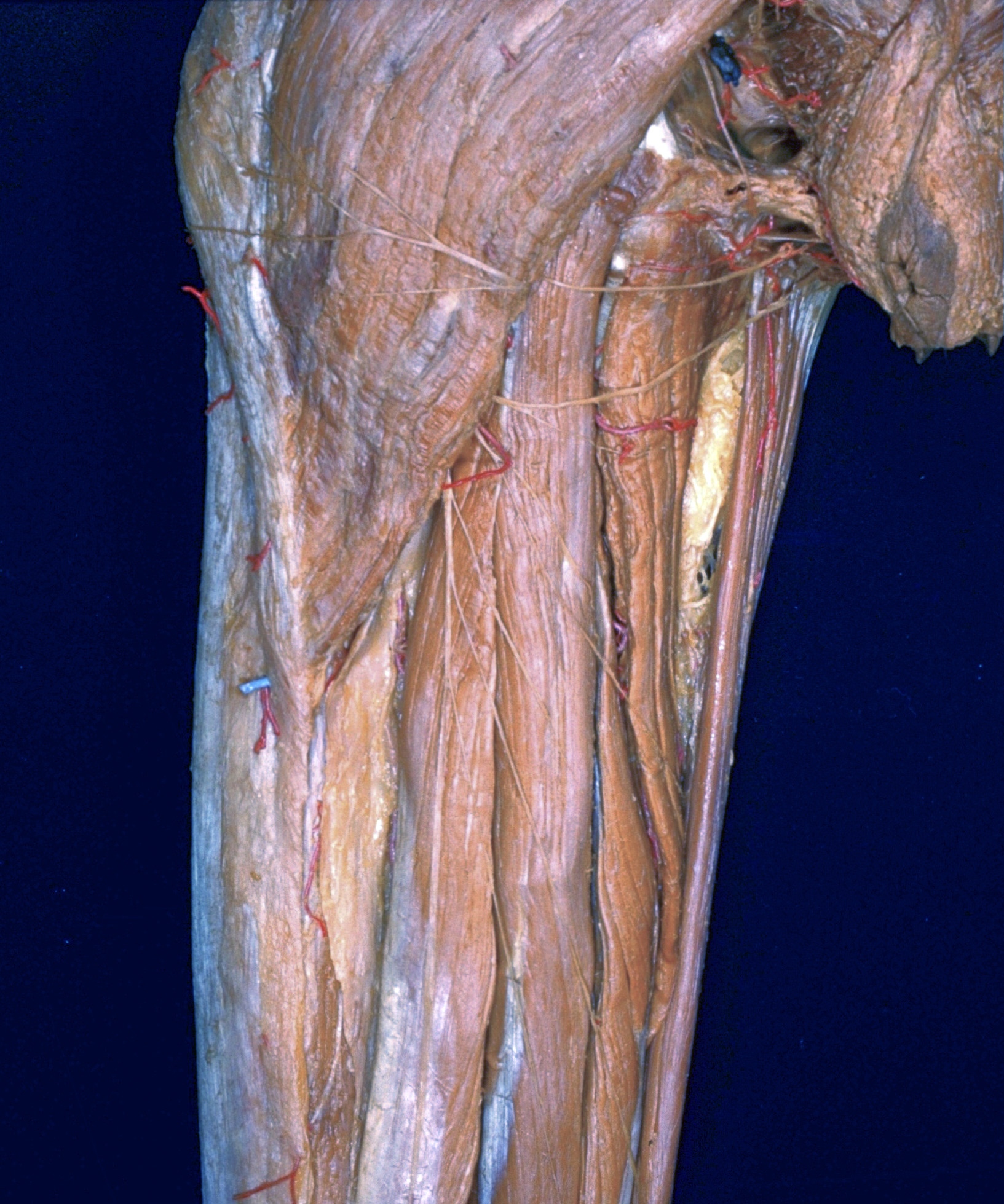
Dissection of posterior aspect of left thigh
Posterior muscles of upper part of thigh, close-up view
Stanford holds the copyright to the David L. Bassett anatomical images and has assigned
Creative Commons license Attribution-Share
Alike 4.0 International to all of the images.
For additional information regarding use and permissions,
please contact the Medical History Center.
Image #182-7
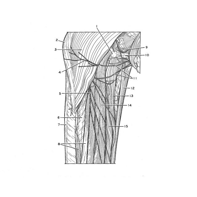
Dissection of posterior aspect of left thigh
Posterior muscles of upper part of thigh, close-up view
The specimen shown in the preceding photograph is illustrated here in a close-up view of the upper thigh.
- Ischial tuberosity
- Position of greater trochanter
- Gluteus maximus muscle
- Inferior cluneal nerve
- Posterior femoral cutaneous nerve (note cutaneous branches passing medially that penetrated fascia lata to reach skin)
- Fascial layer between long and short heads of biceps femoris (not lateral intermuscular septum)
- Iliotibial tract
- Biceps femoris muscle (upper pointer, long head; lower pointer, short head)
- Ischiorectal fossa
- Urogenital diaphragm
- Perineal branch of posterior femoral cutaneous nerve
- Gracilis muscle
- Adductor magnus muscle
- Semitendinosus muscle
- Semimembranosus muscle