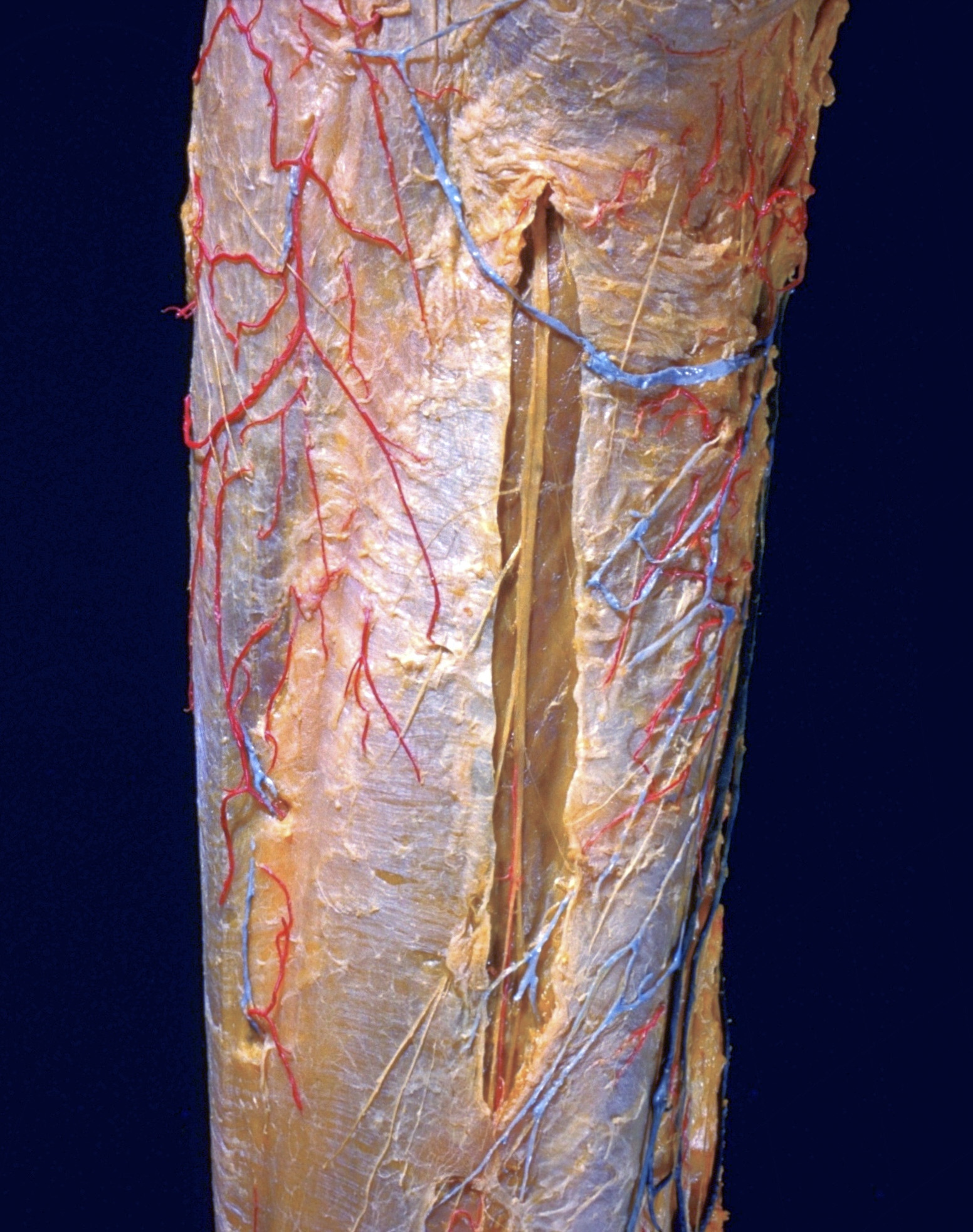
Dissection of posterior aspect of left thigh
Superficial vessels and nerves of mid-thigh, posterior view
Stanford holds the copyright to the David L. Bassett anatomical images and has assigned
Creative Commons license Attribution-Share
Alike 4.0 International to all of the images.
For additional information regarding use and permissions,
please contact the Medical History Center.
Image #182-4
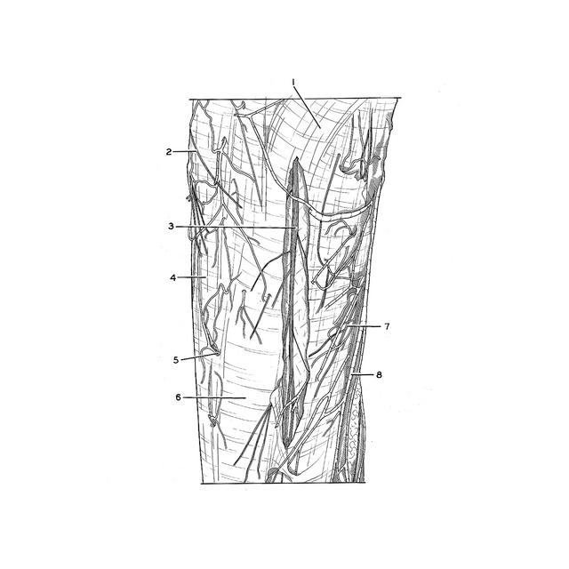
Dissection of posterior aspect of left thigh
Superficial vessels and nerves of mid-thigh, posterior view
This photograph is a close-up view of the specimen which was shown in the previous view. Details of the branching of the posterior femoral cutaneous nerve and of the distribution of branches of other nerves that reach the posterior aspect of the thigh are visible.
- Gluteus maximus muscle (covered by fascia lata)
- Branch of lateral femoral cutaneous nerve
- Posterior femoral cutaneous nerve
- Iliotibial tract (overlying vastus lateralis muscle)
- Branch of perforating artery accompanied by perforating vein
- Biceps femoris muscle (covered by fascia lata)
- Branch of obturator nerve
- Accessory saphenous vein