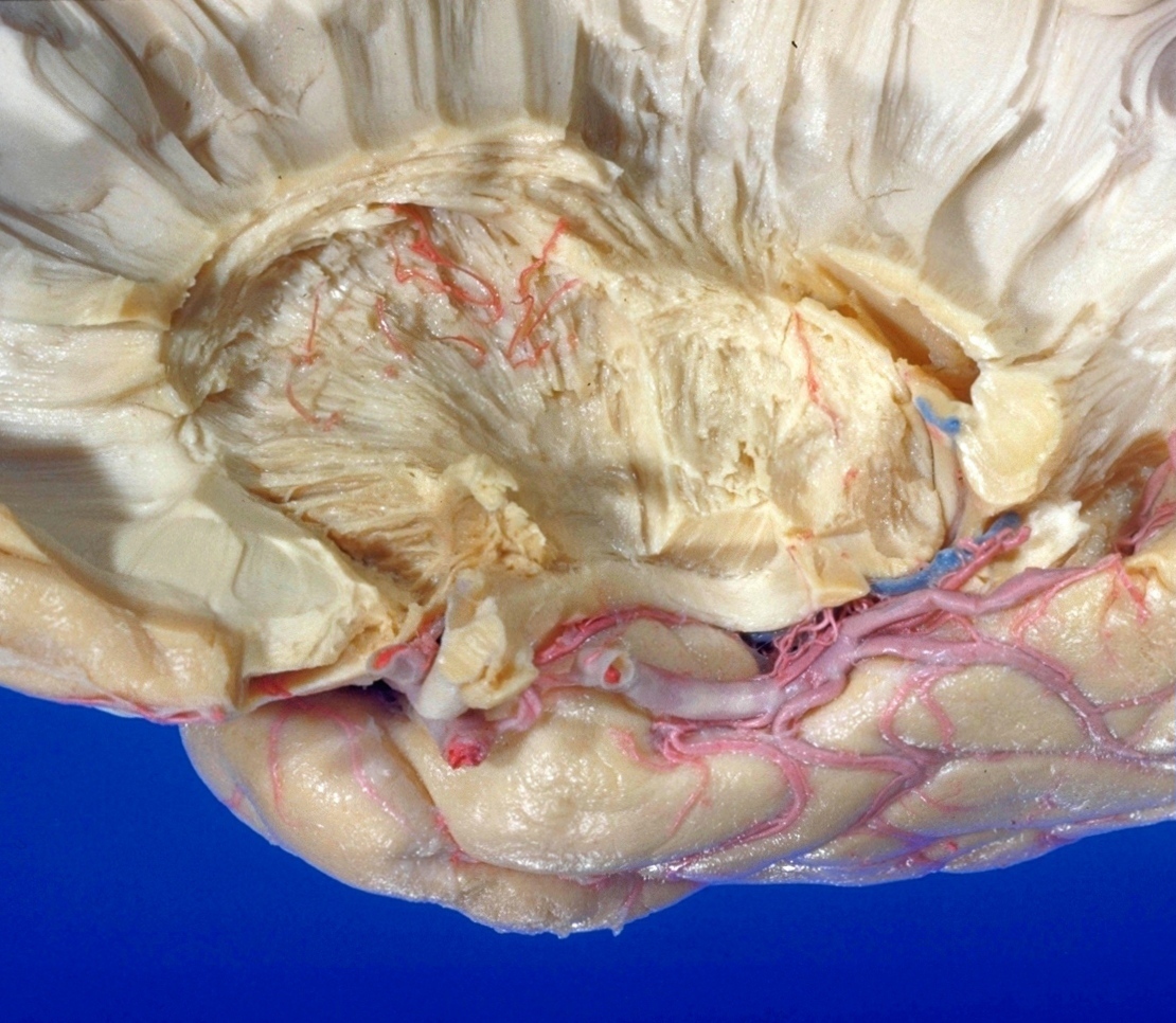
Exploration of the brain from the medial aspect
Lentiform nucleus
Stanford holds the copyright to the David L. Bassett anatomical images and has assigned
Creative Commons license Attribution-Share
Alike 4.0 International to all of the images.
For additional information regarding use and permissions,
please contact the Medical History Center.
Image #17-7
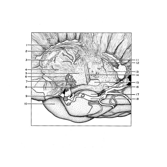
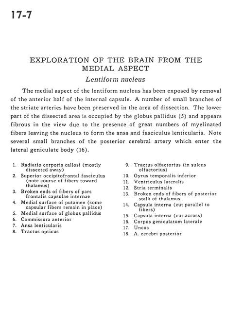
Exploration of the brain from the medial aspect
Lentiform nucleus
The medial aspect of the lentiform nucleus has been exposed by removal of the anterior half of the internal capsule. A number of small branches of the striate arteries have been preserved in the area of dissection. The lower part of the dissected area is occupied by the globus pallidus (5) and appears fibrous in the view due to the presence of great numbers of myelinated fibers leaving the nucleus to form the ansa and fasciculus lenticularis. Note several small branches of the posterior cerebral artery which enter the lateral geniculate body (16).
- Radiation corpus callosum (mostly dissected away)
- Superior occipitofrontal fasciculus (note course of fibers toward thalamus)
- Broken ends of fibers of frontal part internal capsule
- Medial surface of putamen (some capsular fibers remain in place)
- Medial surface of globus pallidus
- Anterior commissure
- Ansa lenticularis
- Optic tract
- Olfactory tract (in olfactory sulcus)
- Inferior temporal gyrus
- Lateral ventricle
- Stria terminalis
- Broken ends of fibers of posterior stalk of thalamus
- Internal capsule (cut parallel to fibers)
- Internal capsule (cut across)
- Lateral geniculate body
- Uncus
- Posterior cerebral artery