Uterus and adnexae
Uterus, uterine tubes and ovaries, anterior aspect
Stanford holds the copyright to the David L. Bassett anatomical images and has assigned
Creative Commons license Attribution-Share
Alike 4.0 International to all of the images.
For additional information regarding use and permissions,
please contact the Medical History Center.
Image #164-2
Uterus and adnexae
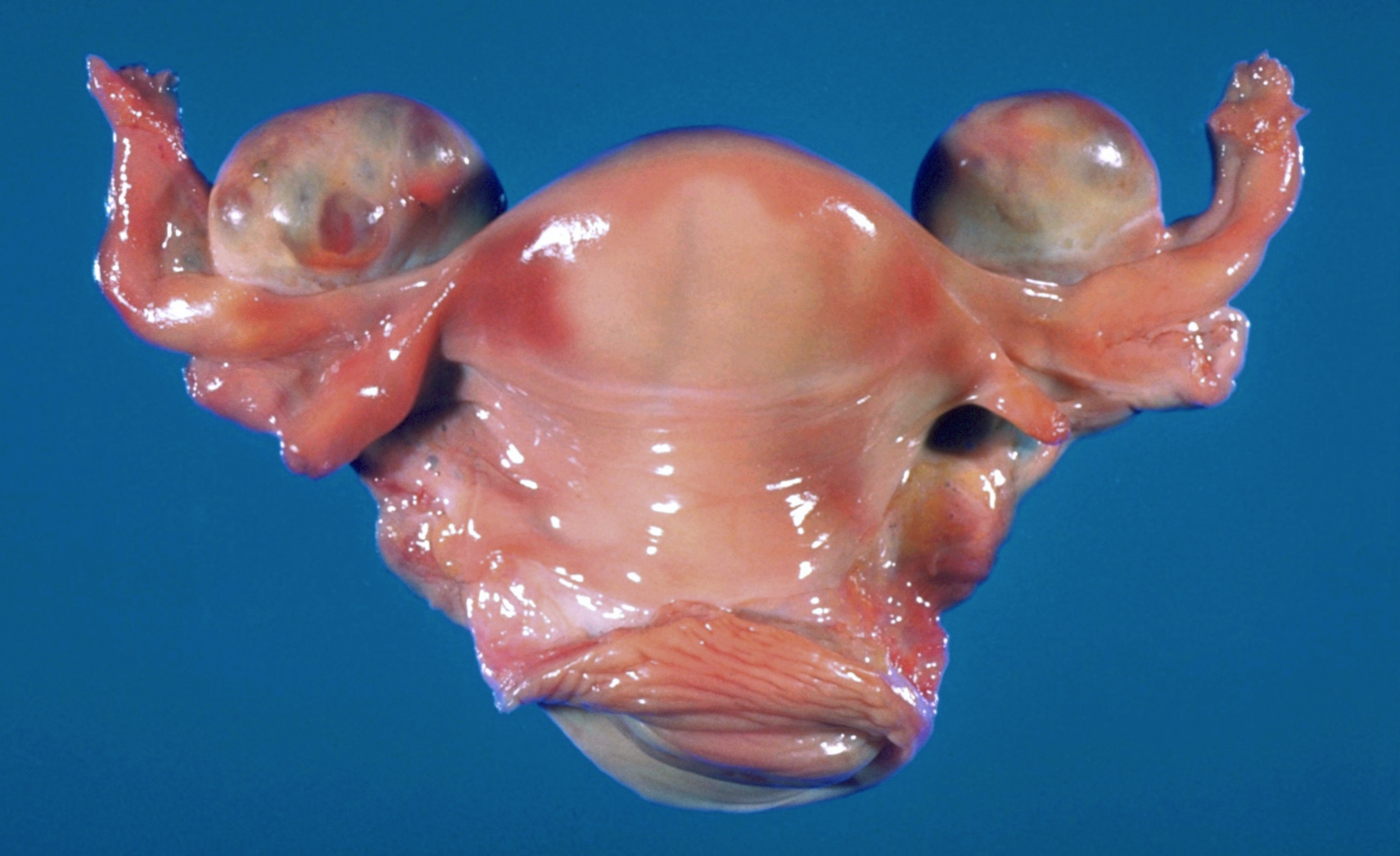
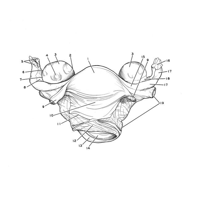
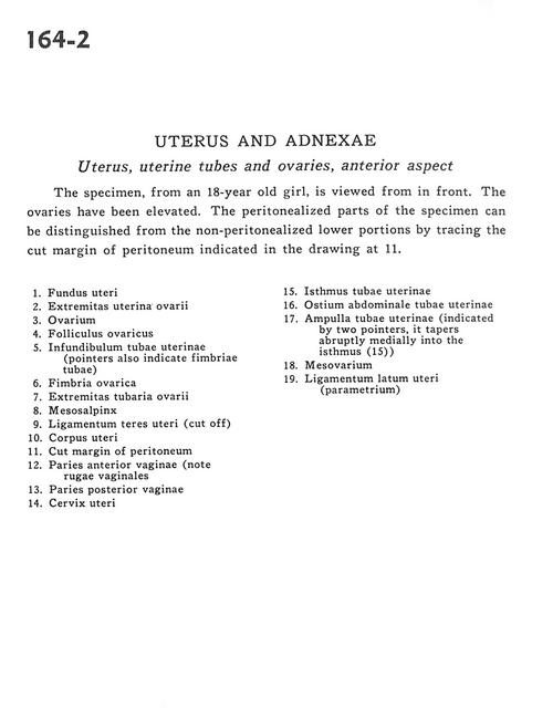
Uterus, uterine tubes and ovaries, anterior aspect
The specimen, from an 18-year old girl, is viewed from in front. The ovaries have been elevated. The peritonealized parts of the specimen can be distinguished from the non-peritonealized lower portions by tracing the cut margin of peritoneum indicated in the drawing at 11.
- Fundus of uterus
- Uterine extremity of ovary
- Ovary
- Ovarian follicle
- Infundibulum of uterine tube (pointers also indicate tubal fimbriae)
- Ovarian fimbria
- Tubal extremity of ovary
- Mesosalpinx
- Ligamentum teres (of uterus) (cut off)
- Body of uterus
- Cut margin of peritoneum
- Anterior wall of vagina (note vaginal rugae)
- Posterior wall of vagina
- Cervix of uterus
- Isthmus of uterine tube
- Abdominal opening of uterine tube
- Ampulla of uterine tube (indicated by two pointers, it tapers abruptly medially into the isthmus (15)
- Mesovarium
- Broad ligament of uterus (parametrium)