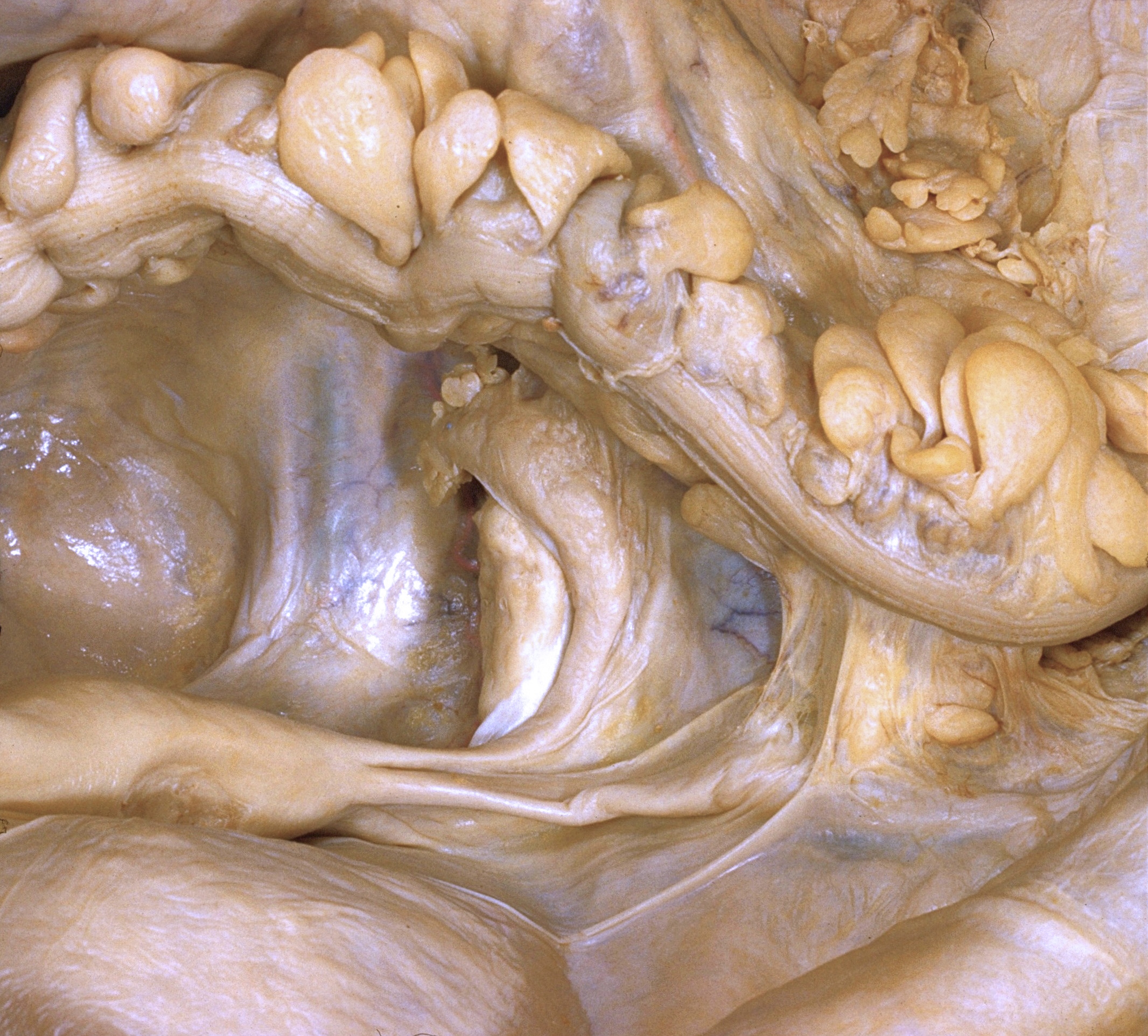
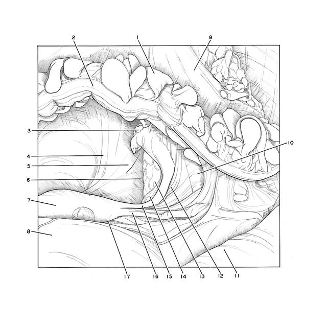
Pelvic peritoneal cavity of female
Left ovarian fossa, close-up view
Stanford holds the copyright to the David L. Bassett anatomical images and has assigned
Creative Commons license Attribution-Share
Alike 4.0 International to all of the images.
For additional information regarding use and permissions,
please contact the Medical History Center.
Image #164-1
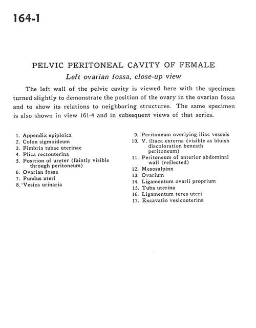
Pelvic peritoneal cavity of female
Left ovarian fossa, close-up view
The left wall of the pelvic cavity is viewed here with the specimen turned slightly to demonstrate the position of the ovary in the ovarian fossa and to show its relations to neighboring structures. The same specimen is also shown in view 161-4 and in subsequent views of that series.
- Appendix epiploica
- Sigmoid colon
- Fimbria of uterine tube
- Rectouterine fold
- Position of ureter (faintly visible through peritoneum)
- Ovarian fossa
- Fundus of uterus
- Urinary bladder
- Peritoneum overlying iliac vessels
- External iliac vein (visible as bluish discoloration beneath peritoneum)
- Peritoneum of anterior abdominal wall (reflected)
- Mesosalpinx
- Ovary
- Proper ovarian ligament
- Uterine tube
- Ligamentum teres (of uterus)
- Uterovesical pouch