Dissection of female pelvis from a lateral approach
Pelvic plexus and ganglion; vaginal artery
Stanford holds the copyright to the David L. Bassett anatomical images and has assigned
Creative Commons license Attribution-Share
Alike 4.0 International to all of the images.
For additional information regarding use and permissions,
please contact the Medical History Center.
Image #162-6
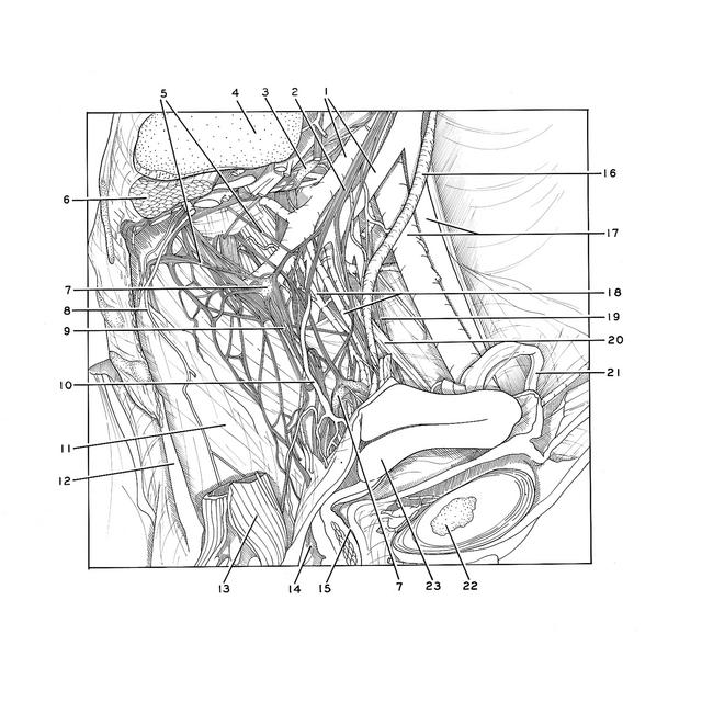

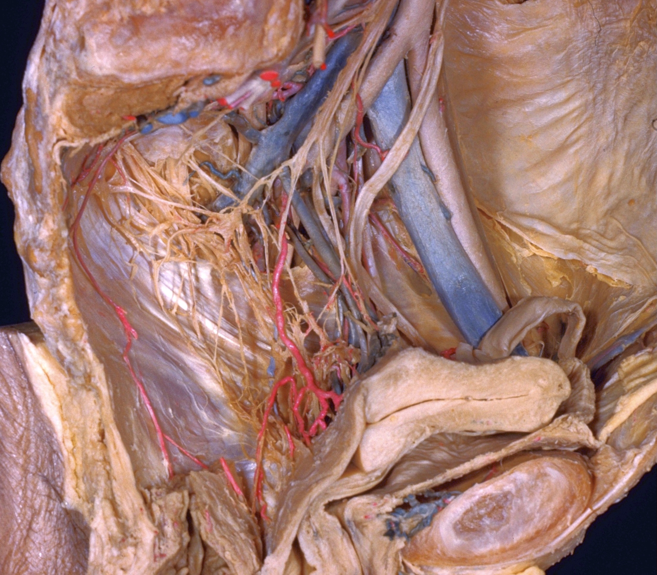
Dissection of female pelvis from a lateral approach
Pelvic plexus and ganglion; vaginal artery
The uterus and vagina have now been deflected anteriorly. The uterosacral and cardinal ligaments, which are shown in 162-2, have been dissected so that the pelvic plexus and the vaginal and uterine blood vessels are exposed.
- Internal iliac artery and vein
- Superior hypogastric plexus
- Ganglion of sympathetic trunk
- Articular surface of sacrum
- Pelvic splanchnic nerves
- Piriform muscle (cut across)
- Pelvic ganglion
- Middle sacral artery
- Inferior hypogastric plexus (pelvic plexus)
- Vaginal artery (branch of uterine artery)
- Superior fascia of pelvic diaphragm
- Pelvic diaphragm (sectioned near midline)
- Rectum
- Vagina
- Urethra
- Ureter
- External iliac artery and vein
- Uterine veins
- Obturator nerve
- Lateral umbilical ligament
- Ligamentum teres (of uterus)
- Pubic symphysis
- Uterus