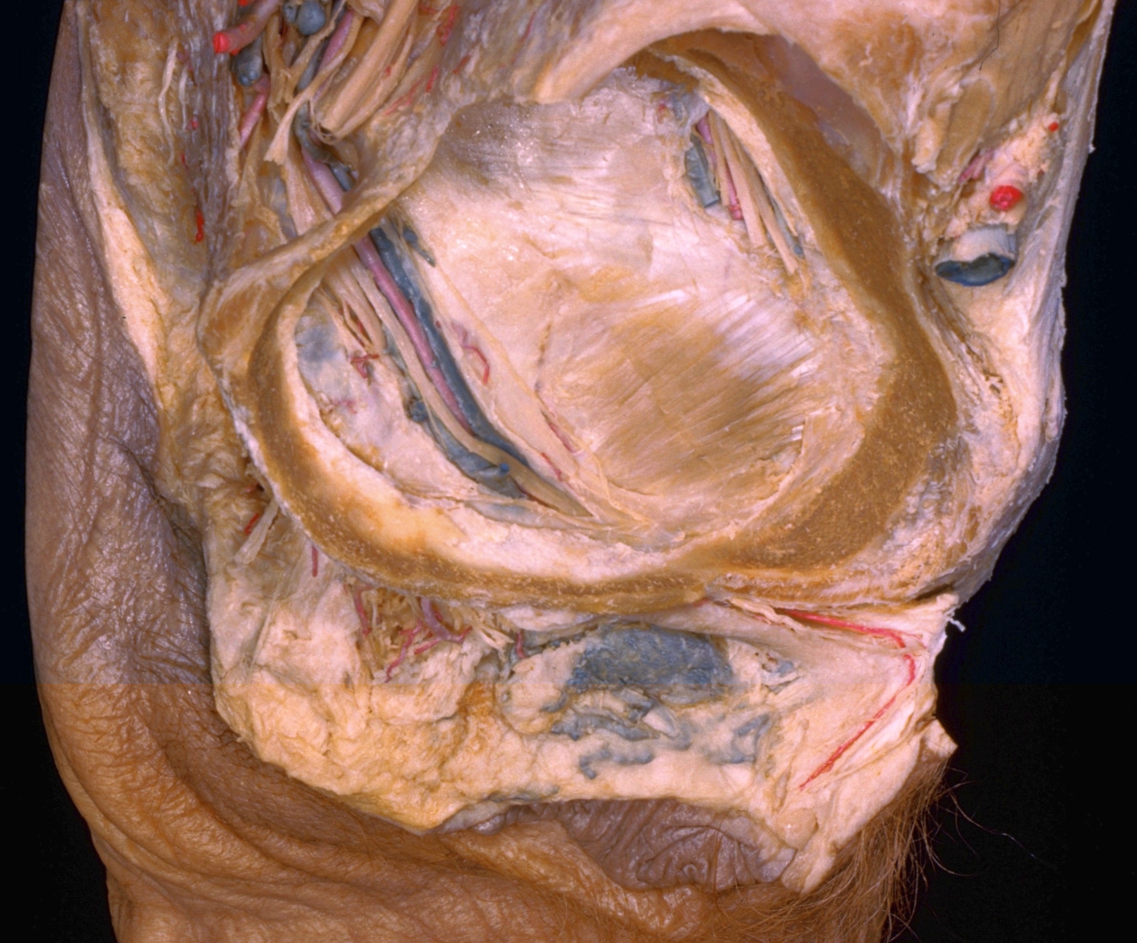
Dissection of female pelvis froma lateral approach
Pudendal nerve and branches in situ
Stanford holds the copyright to the David L. Bassett anatomical images and has assigned
Creative Commons license Attribution-Share
Alike 4.0 International to all of the images.
For additional information regarding use and permissions,
please contact the Medical History Center.
Image #159-2
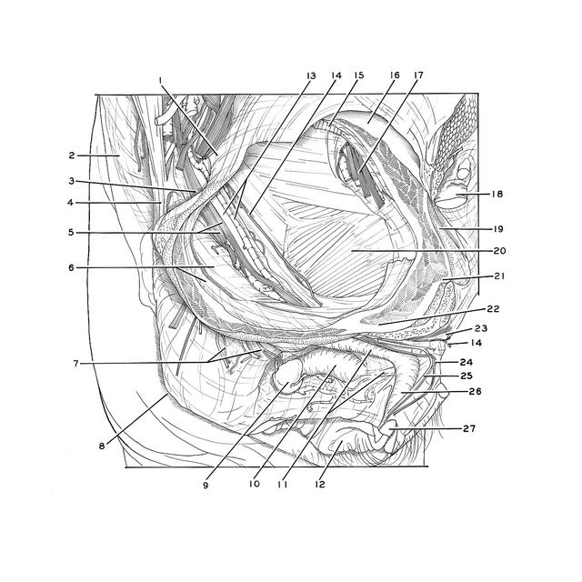
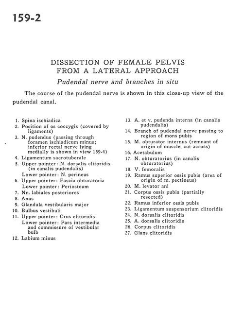
Dissection of female pelvis froma lateral approach
Pudendal nerve and branches in situ
The course of the pudendal nerve is shown in this close-up view of the pudendal canal.
- Ischial spine
- Position of coccyx (covered by ligaments)
- Pudendal nerve (passing through lesser sciatic foramen; inferior rectal nerve lying medially is shown in view 159-4)
- Sacrotuberous ligament
- Upper pointer: Dorsal nerve of clitoris (in pudendal canal) Lower pointer: Perineal nerve
- Upper pointer: Obturator fascia Lower pointer: Periosteum
- Posterior labial nerves
- Anus
- Major vestibular gland
- Vestibular bulb
- Upper pointer: Crus of clitoris Lower pointer: Intermediate part and commissure of vestibular bulb
- Labium minus
- Internal pudendal artery and vein (in pudendal canal)
- Branch of pudendal nerve passing to region of mons pubis
- Obturator internus muscle (remnant of origin of muscle, cut across)
- Acetabulum
- Obturator nerve (in obturator canal)
- Femoral vein
- Superior pubic ramus (area of origin of pectineus muscle)
- Levator ani muscle
- Body of pubic bone (partially resected)
- Inferior pubic ramus
- Suspensory ligament of clitoris
- Dorsal nerve of clitoris
- Dorsal artery of clitoris
- Body of clitoris
- Glans of clitoris