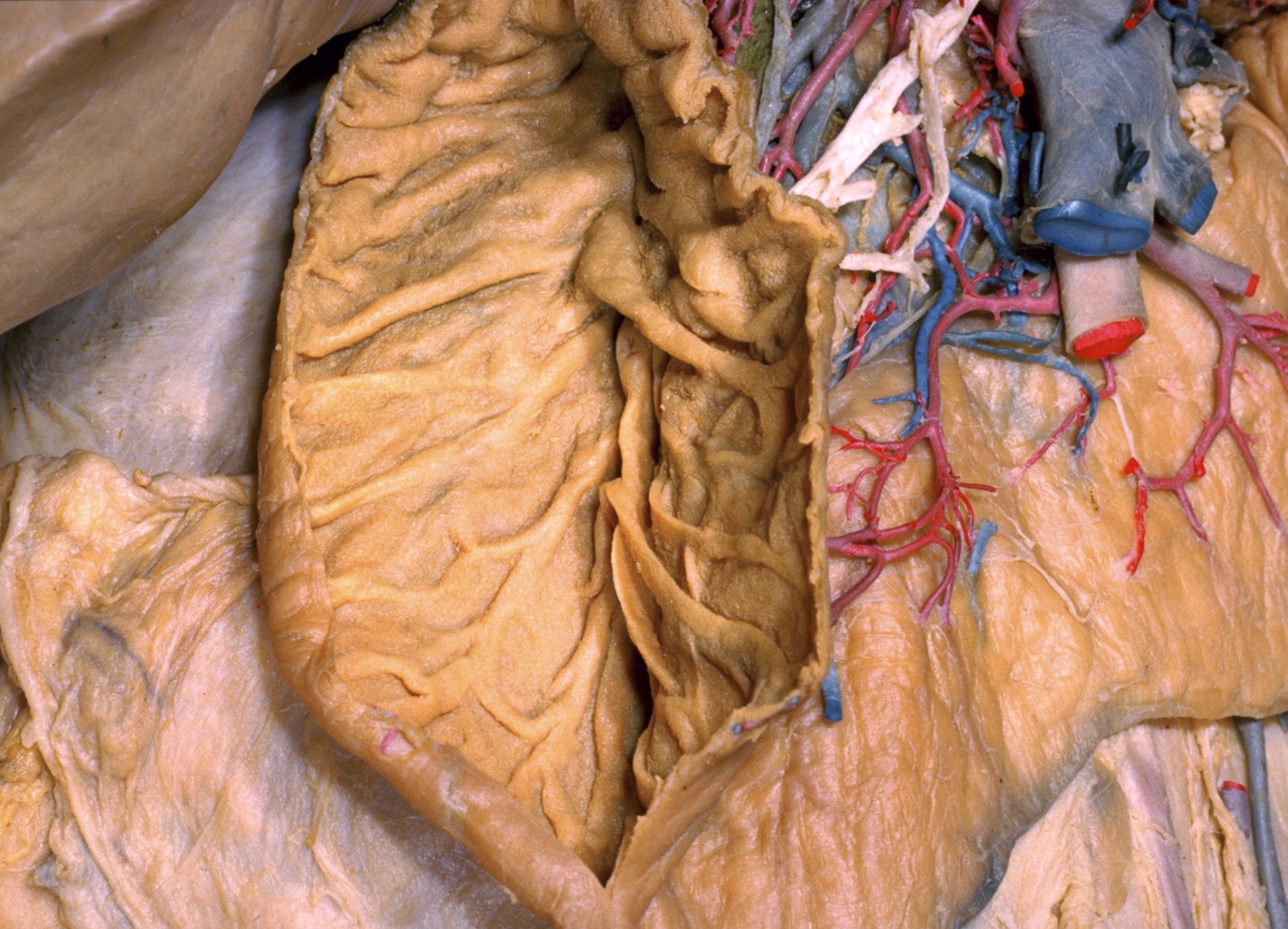
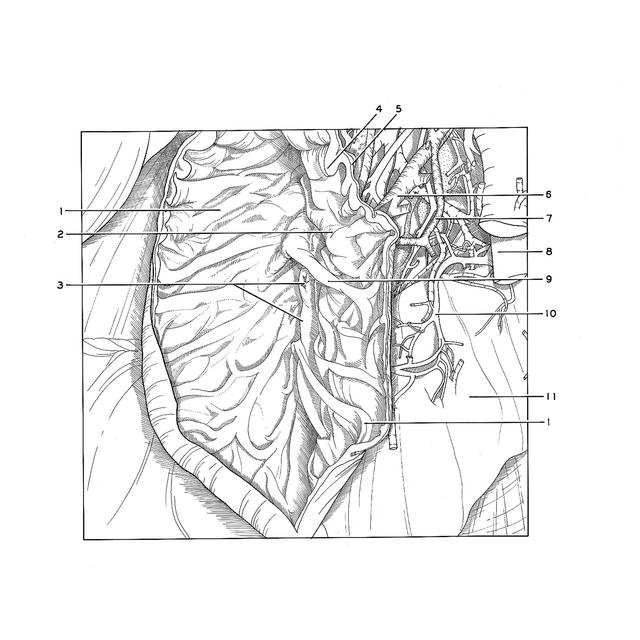
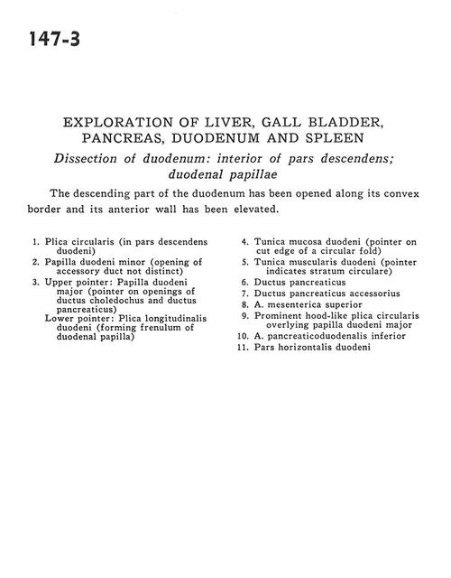
Exploration of liver, gall bladder, pancreas, duodenum and spleen
Dissection of duodenum; interior of pars descendens; duodenal papillae
Stanford holds the copyright to the David L. Bassett anatomical images and has assigned
Creative Commons license Attribution-Share
Alike 4.0 International to all of the images.
For additional information regarding use and permissions,
please contact the Medical History Center.
Image #147-3
Exploration of liver, gall bladder, pancreas, duodenum and spleen
Dissection of duodenum; interior of pars descendens; duodenal papillae
The descending part of the duodenum has been opened along its convex border and its anterior wall has been elevated.
- Plica circularis (in descending part of duodenum)
- Minor duodenal papilla (opening of accessory duct not distinct)
- Upper pointer: Major duodenal papilla (pointer on openings of common bile duct and pancreatic duct) Lower pointer: Longitudinal duodenal fold (forming frenulum of duodenal papilla)
- Muscular layer of duodenum (pointer on cut edge of a circular fold)
- Muscular layer of duodenum (pointer indicates circular fibers)
- Pancreatic duct
- Accessory pancreatic duct
- Superior mesenteric artery
- Prominent hood-like plica circularis overlying major duodenal papilla
- Inferior pancreaticoduodenal artery
- Horizontal part of duodenum