Dissection of thorax from a posterior approach
Ligaments of costotransverse articulations in mid-thoracic region
Stanford holds the copyright to the David L. Bassett anatomical images and has assigned
Creative Commons license Attribution-Share
Alike 4.0 International to all of the images.
For additional information regarding use and permissions,
please contact the Medical History Center.
Image #131-6
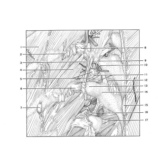
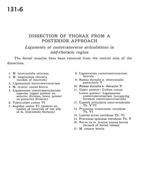
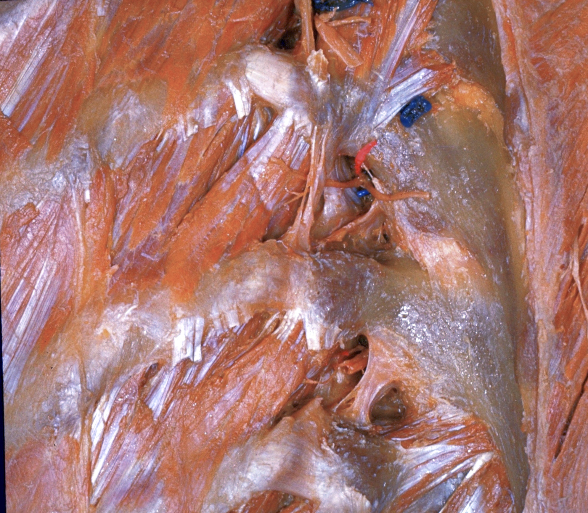
Dissection of thorax from a posterior approach
Ligaments of costotransverse articulations in mid-thoracic region
The dorsal muscles have been removed from the central area of the dissection.
- External intercostal muscle
- Longissimus thoracis muscle (tendon of insertion)
- Intertransverse ligament
- Levator costarum brevis muscle
- Superior costotransverse ligament (upper pointer on anterior division, lower pointer on posterior division)
- Costal tubercle VI
- Angle of rib VI (pointer on tendon of insertion of one slip of iliocostalis thoracis muscle)
- Lateral costotransverse ligament
- Dorsal branch posterior intercostal artery V
- Dorsal branch thoracic nerve V
- Upper pointer: Neck of rib Lower pointer: Costotransverse ligament (occupying foramen costotransversarium)
- Intervertebral joint capsule Th. V-VI
- Transverse process Th. vertebra VI
- Lamina of vertebral arch Th. VI
- Spinous process vertebrae Th. VI
- Nerve to levator costarum brevis muscle (branch of dorsal branch)
- Rotator brevis muscle