Dissection of thorax from a posterior approach
Deep musculature.
Stanford holds the copyright to the David L. Bassett anatomical images and has assigned
Creative Commons license Attribution-Share
Alike 4.0 International to all of the images.
For additional information regarding use and permissions,
please contact the Medical History Center.
Image #131-2
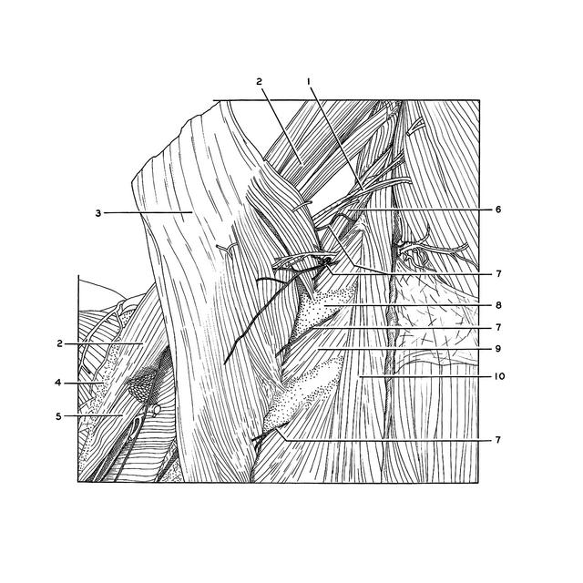
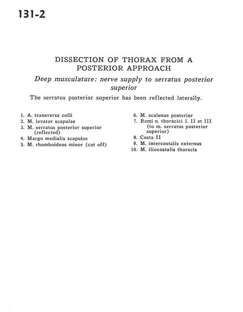
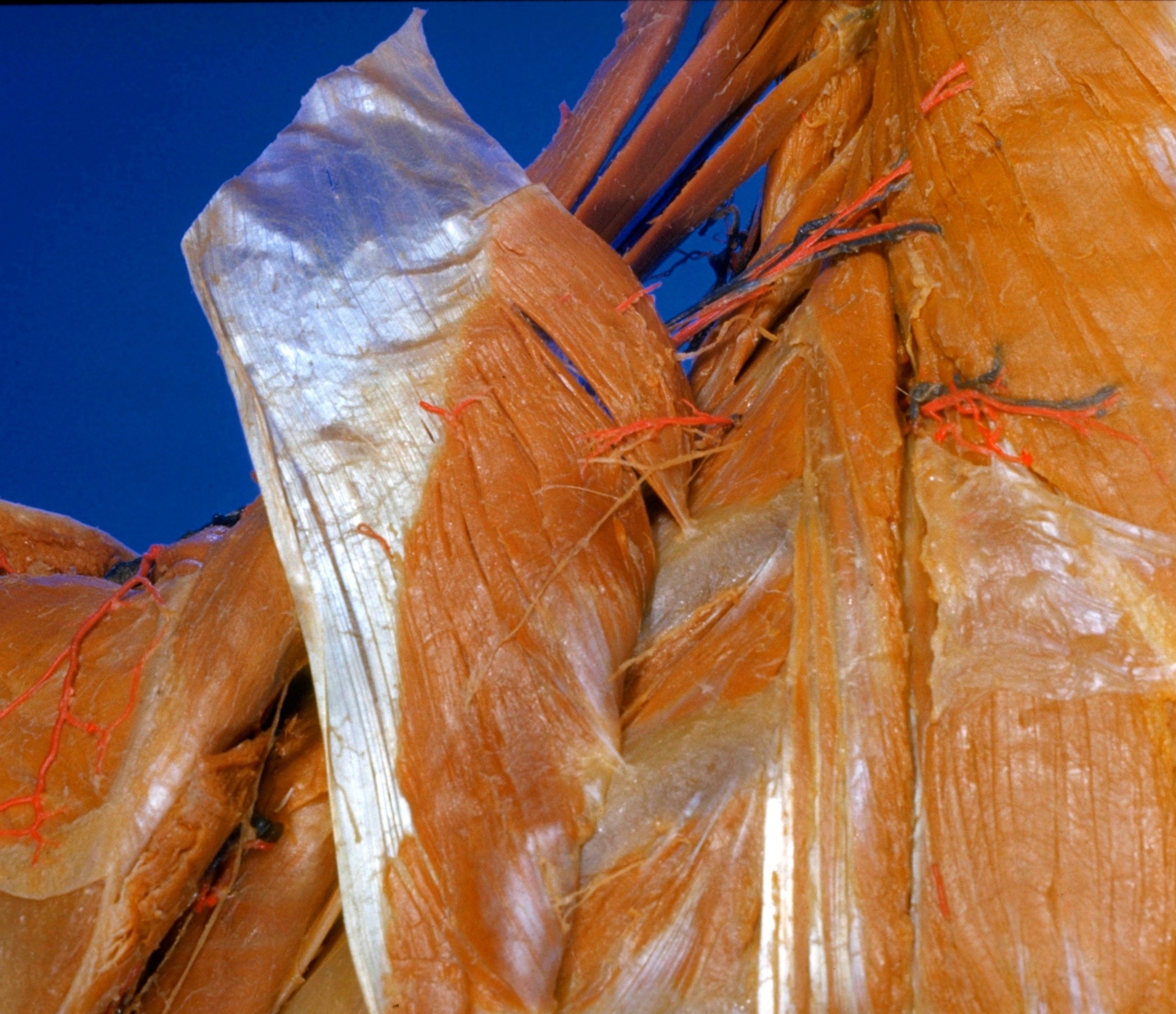
Dissection of thorax from a posterior approach
Deep musculature.
The serratus posterior superior has been reflected laterally.
- Transverse colli artery
- Levator scapulae muscle
- Serratus posterior superior muscle (reflected)
- Medial scapular margin
- Rhomboid minor muscle (cut off)
- Posterior scalene muscle
- Branches of thoracic nerve I, II and III (to serratus posterior superior muscle)
- Rib II
- External intercostal muscle
- Iliocostalis thoracis muscle